2010 Winter Project Week TBISegmentation
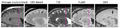
TBI modalities
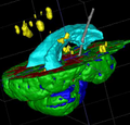
Segmentation result with lesion and shunt


Key Investigators
- Marcel Prastawa, Guido Gerig, University of Utah
- Ron Kikinis, BWH
- UCLA
Objective
Traumatic Brain Injury (TBI) is caused by severe impact to the brain, which may result in skull fracture, lesions, and internal bleeding. Full assessment of these injuries is possible via multi-modality imaging, here T1w, T2, T1-postcontrast, Flair (fluid attenuated inversion recovery), SWI (susceptibility weighted imaging), DTI). Joint analysis of these modalities requires co-registration of these sets which come with different orientations, spatial resolution and head coverage. Segmentation of brain tissue, fluid and pathology requires efficient procedures for multi-modal analysis of images with classification of lesions.
Approach, Plan
Preferably, such a procedure should be automatic given the presence of small-scale pathology such as lesions, bleedings and ventricular shape alterations, or involve efficient, easy and intuitive expert interaction to support an automated classification algorithm. We propose an atlas based multi-modal segmentation method (ABC: Atlas-Based Classification), which makes use of normative data (spatial and intensity) for isolating abnormal regions that are likely due to injury.
Progress
We have a working ptototype program of ABC written in C++ using ITK. ABC incorporates image co-registration, atlas template registration, bias field correction, and tissue classification into an efficient workflow. We will wrap it as a Slicer module and will systematically test its performance on various TBI datasets. These tests will include co-registration of DTI and structural MRI for joint analysis of white matter tracts, brain anatomy and lesions.