Difference between revisions of "RSNA Quantitative Imaging Course"
m (Text replacement - "http://www.slicer.org/slicerWiki/index.php/" to "https://www.slicer.org/wiki/") |
|||
| (3 intermediate revisions by one other user not shown) | |||
| Line 1: | Line 1: | ||
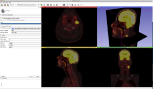
[[image:Screen_Shot_2012-11-20_at_12.10.39_AM.png |right|300px]] | [[image:Screen_Shot_2012-11-20_at_12.10.39_AM.png |right|300px]] | ||
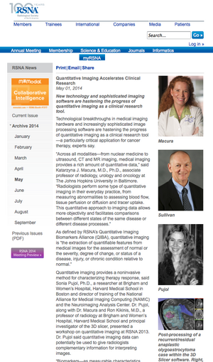
[[Image:RSNAInterview_PujolMacura.png |right|300 px]] | [[Image:RSNAInterview_PujolMacura.png |right|300 px]] | ||
| − | Technological breakthroughs in medical imaging hardware and the emergence of increasingly sophisticated image processing software tools permit the visualization and display of complex anatomical structures with increasing sensitivity and specificity. This course begins with an introductory presentation of state-of-the-art, clinical examples of quantitative imaging biomarkers for diagnosis and clinical trial outcome measures. Cases from multiple imaging modalities and from multiple organ systems will be highlighted to illustrate the depth and breath of this field. Participants will then be led through a series of tutorials on the basics of viewing and processing DICOM volumes in 3D using [http://www.slicer.org 3D Slicer]. A | + | Technological breakthroughs in medical imaging hardware and the emergence of increasingly sophisticated image processing software tools permit the visualization and display of complex anatomical structures with increasing sensitivity and specificity. This course begins with an introductory presentation of state-of-the-art, clinical examples of quantitative imaging biomarkers for diagnosis and clinical trial outcome measures. Cases from multiple imaging modalities and from multiple organ systems will be highlighted to illustrate the depth and breath of this field. Participants will then be led through a series of tutorials on the basics of viewing and processing DICOM volumes in 3D using [http://www.slicer.org 3D Slicer]. A [[media:QuantitativeImaging_SoniaPujol_RSNA2014_Dec2.pdf | Quantitative Imaging tutorial ]] with pre-computed datasets will focus on basic use of 3D Slicer software, quantitative measurements from PET/CT studies, and volumetric analysis of meningioma. |
This [[RSNA_Quantitative_Imaging_Course |course]] is offered in collaboration with the Johns Hopkins Institute for Clinical and Translational Research. | This [[RSNA_Quantitative_Imaging_Course |course]] is offered in collaboration with the Johns Hopkins Institute for Clinical and Translational Research. | ||
| − | For additional training materials on the software, please visit the [ | + | For additional training materials on the software, please visit the [https://www.slicer.org/wiki/Documentation/4.3/Training 3D Slicer Compendium]. [http://www.slicer.org/slicerWiki/images/4/46/QuantitativeImaging.zip Course datasets available here] |
The RSNA quantitative Imaging Course was featured in [http://rsna.org/NewsDetail.aspx?id=12002 RSNA News]. | The RSNA quantitative Imaging Course was featured in [http://rsna.org/NewsDetail.aspx?id=12002 RSNA News]. | ||
Latest revision as of 17:29, 10 July 2017
Home < RSNA Quantitative Imaging CourseTechnological breakthroughs in medical imaging hardware and the emergence of increasingly sophisticated image processing software tools permit the visualization and display of complex anatomical structures with increasing sensitivity and specificity. This course begins with an introductory presentation of state-of-the-art, clinical examples of quantitative imaging biomarkers for diagnosis and clinical trial outcome measures. Cases from multiple imaging modalities and from multiple organ systems will be highlighted to illustrate the depth and breath of this field. Participants will then be led through a series of tutorials on the basics of viewing and processing DICOM volumes in 3D using 3D Slicer. A Quantitative Imaging tutorial with pre-computed datasets will focus on basic use of 3D Slicer software, quantitative measurements from PET/CT studies, and volumetric analysis of meningioma. This course is offered in collaboration with the Johns Hopkins Institute for Clinical and Translational Research. For additional training materials on the software, please visit the 3D Slicer Compendium. Course datasets available here
The RSNA quantitative Imaging Course was featured in RSNA News.
- CME Credit Categories: Biomarkers/Quantitative Imaging; Informatics
- AMA PRA Category 1 Credits™: 1.5
- ARRT Category A+ Credit: 1.5
Teaching Faculty
- Sonia Pujol, Ph.D., Surgical Planning Laboratory, Department of Radiology, Brigham and Women’s Hospital, Harvard Medical School, Boston MA.
- Katarzyna J. Macura MD, PhD., Department of Radiology and Radiological Sciences, The Johns Hopkins Medical Institutions, Baltimore, MD.
- Ron Kikinis, M.D., Surgical Planning Laboratory, Department of Radiology, Brigham and Women’s Hospital, Harvard Medical School, Boston MA.
Logistics
- Date: Tuesday December 2, 8:30-10 am
- Location: Room S401AB, McCormick Conference Center, Chicago, IL.
- Registration: Available through the RSNA 2014 website.

