Difference between revisions of "2017 Winter Project Week/Knee Cartilage Segmentation"
From NAMIC Wiki
Hans.meine (talk | contribs) |
Hans.meine (talk | contribs) |
||
| Line 56: | Line 56: | ||
=== Planned Visualization === | === Planned Visualization === | ||
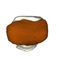
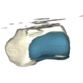
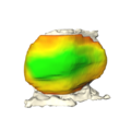
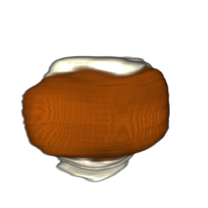
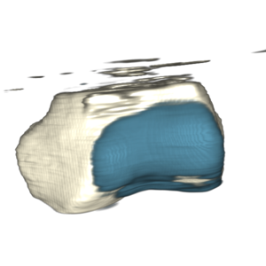
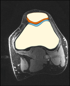
[[File:Patella_snapshot.png|300px|3d rendering of patella with cartilage]][[File:Femur_snapshot.png|300px|3d rendering of femur with cartilage]][[File:Ortho_snapshot.png|245px|2d slice of MRI with segmented structures]] | [[File:Patella_snapshot.png|300px|3d rendering of patella with cartilage]][[File:Femur_snapshot.png|300px|3d rendering of femur with cartilage]][[File:Ortho_snapshot.png|245px|2d slice of MRI with segmented structures]] | ||
| + | |||
Below this should've been the performance over time, but with a rendering of the increasing number of training cases (1..47). | Below this should've been the performance over time, but with a rendering of the increasing number of training cases (1..47). | ||
Latest revision as of 15:29, 13 January 2017
Home < 2017 Winter Project Week < Knee Cartilage SegmentationKey Investigators
- Hans Meine (University of Bremen, MEVIS)
Project Description
| Objective | Approach and Plan | Progress and Next Steps |
|---|---|---|
|
|
|
Background and References
Goal / context
- analysis of cartilage thickness for ostheoarthitis assessment
- High-resolution data is from the University of Freiburg (T. Lange, K. Izadpanah)
- patellofemoral joint (prospective motion correction)
- depicted image is a healthy volunteer
- cf. CARS 2016 submission
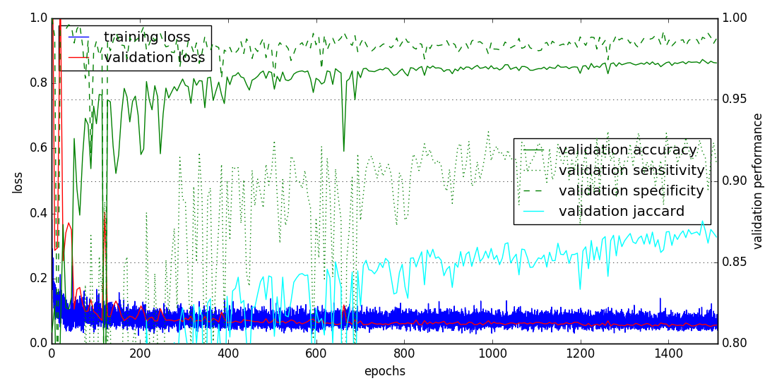
Training worked fine
Planned Visualization
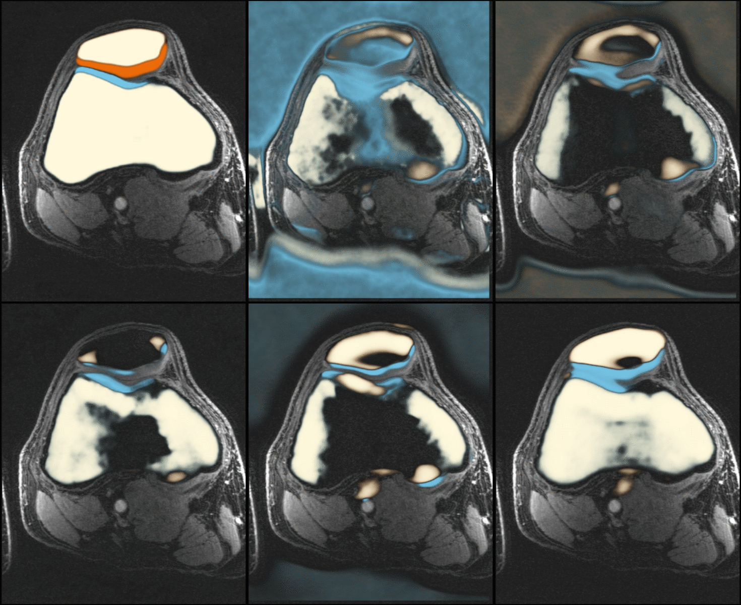
Below this should've been the performance over time, but with a rendering of the increasing number of training cases (1..47).
Glimpse at partial animation
This is only the 2d part, I cancelled the rendering because the 3d cameras were not properly set up, and made a mistake, completely shutting myself out of remote access to the machine required for this project. :-(


