Difference between revisions of "2012 Summer Project Week:Interactive Needle Segmentation"
From NAMIC Wiki
| Line 4: | Line 4: | ||
Image:PW-MIT2012.png|[[2012_Summer_Project_Week#Projects|Projects List]] | Image:PW-MIT2012.png|[[2012_Summer_Project_Week#Projects|Projects List]] | ||
| − | Image: | + | Image:gyn-needle-segmentation-1.png|Screenshot of Manual Needle Segmentation (20 minutes) |
| − | |||
| − | |||
</gallery> | </gallery> | ||
Revision as of 04:41, 18 June 2012
Home < 2012 Summer Project Week:Interactive Needle Segmentation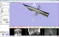
Screenshot of Manual Needle Segmentation (20 minutes)


Key Investigators
- Rutgers: Nabgha Farhat
- SPL: Nabgha Farhat, Jan Egger, Tina Kapur, Steve Pieper
Objective
The goal of this project is to achieve fast (< 2 minute) interactive segmentation of 5-10 needles from MRI images using Slicer.
Approach, Plan
We will explore different segmentation/editing tools in Slicer to determine which ones can most efficiently segment 2mm needles (10-15 cm long), from MRI scans in which voxels are about 2mm in each dimension. (These needles are not straight lines, and are often placed within a few mm of each other.)
Progress
Delivery Mechanism
N/A