Difference between revisions of "Events: DTI Tractography Challenge"
From NAMIC Wiki
| Line 30: | Line 30: | ||
* Yundi Shi, UNC Chapell Hill, USA | * Yundi Shi, UNC Chapell Hill, USA | ||
| − | == Corticospinal tract == | + | == Corticospinal tract definition == |
| − | + | ||
| + | {| border="1" cellpadding="5" cellspacing="0" align="center" | ||
| + | | colspan="3" align="center" | | ||
| + | {| border="0" | ||
| + | |+|- | ||
| + | | align="center" width="300px" |[[Image:Corticospinaltract.bmp|350px|thumb|Corticospinal tract. Source: Barrett KE, Barman SM, Boitano S, Brooks H. Ganong's Review of Medical Physiology, 23rd Edition. http://www.accessmedicine.com]] | ||
| + | | align="center" width="300px" |[[File:BrodmannAreas.bmp |thumb|350px|right|Lateral aspect of the cerebrum. The cortical areas are shown according to Brodmann with functional localizations. Source: Waxman SG. Clinical Neuroanatomy,26th Edition. http://www.accessmedicine.com]] | ||
| + | | align="center" width="300px" |[[File:MotorHomunculus.bmp|thumb|250px|right|Motor Homunculus. Source: Penfield W. and Rasmussen T. The Cerebral Cortex of Man, New York, Macmillan, 1950.]] | ||
| + | |- | ||
| + | |} | ||
| + | |} | ||
| + | |||
<gallery widths=300px heights=300px perrow=3 caption="Cortico Spinal Tract (CST), from the 2008 edition of the PNL SPL brain atlas"> | <gallery widths=300px heights=300px perrow=3 caption="Cortico Spinal Tract (CST), from the 2008 edition of the PNL SPL brain atlas"> | ||
image:CST-SPL-PNL-Atlas-2008-Issue.png|CST and surrounding structures: Putamen blue, caudate nucleus in brown, lateral ventricles in green | image:CST-SPL-PNL-Atlas-2008-Issue.png|CST and surrounding structures: Putamen blue, caudate nucleus in brown, lateral ventricles in green | ||
Revision as of 05:02, 23 September 2011
Home < Events: DTI Tractography ChallengeThe DTI Challenge workshop was held at the MICCAI 2011 conference in Toronto, Canada. Please visit the workshop page of the event for details on the organization.
This page will gather material on the on-going effort and workshop outcomes.
(Page under construction)
Workshop Faculty
- Sonia Pujol, Ph.D., Surgical Planning Laboratory, Brigham and Women’s Hospital, Harvard Medical School
- Ron Kikinis, M.D., Surgical Planning Laboratory, Brigham and Women’s Hospital, Harvard Medical School
- Alexandra Golby, M.D., Department of Neurosurgery, Brigham and Women’s Hospital, Harvard Medical School
- Guido Gerig, Ph.D.,The Scientific Computing and Imaging Institute, University of Utah
- Martin Styner, Ph.D., Neuro Image Research and Analysis Laboratory, University of North Carolina
- William Wells, Ph.D., Surgical Planning Laboratory, Brigham and Women’s Hospital, Harvard Medical School
- Carl-Fredrik Westin, Ph.D., Laboratory of Mathematics in Imaging, Brigham and Women’s Hospital, Harvard Medical School
- Sylvain Gouttard, M.Sc.,The Scientific Computing and Imaging Institute, University of Utah
- Arya Nabavi, M.D., Department of Neurosurgery, University Hospital Schleswig-Holstein, Kiel, Germany
Teams participating in the challenge
- Olivier Commonwick, INRIA Rennes, France
- Caroline Brun, UPenn, Philadelphia, USA
- Alonso Ramirez, Ramon Aranda, University of Guanajuato, Mexico
- Maged Goubran, Robarts Research Institute, Toronto, Canada
- Gopal Veni, Scientific Computing and Imaging Institute, Salt Lake City, USA
- Guang Cheng, University of Florida, USA
- Antonio Tristan-Vega, Laboratory of Mathematics in Imaging, Boston, USA
- Peter Neher, German Cancer Research Centre, Heidelberg, Germany
- Yundi Shi, UNC Chapell Hill, USA
Corticospinal tract definition
| |||||


- Cortico Spinal Tract (CST), from the 2008 edition of the PNL SPL brain atlas

CST and surrounding structures: Putamen blue, caudate nucleus in brown, lateral ventricles in green
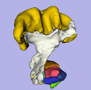
3d rendering of the CST. Motor Cortex in Yellow, CST in white, thalamic nuclei in multiple colors
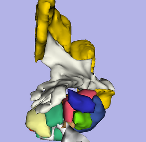
3d rendering of the CST, view from below


- The atlas is freely available. For a download, see here. In addition to the label maps, it is configured to be loaded into 3D Slicer. Several preconfigured views are available.
- For more information about where the CST is located in the posterior limb in the internal capsule and spinal cord, have a look at the picture in this link.