Home < 2017 Winter Project Week < HyperspectralOpht
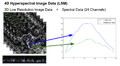
3D hrSIM image data and 4D LSM data
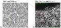
Cell segmentation on 3D SIM data. The segmentation mask is overlayed on 4D LSM data for spectral analysis.
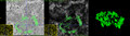
Individual granules (Lipofuscin (bright) and Melano-lipofuscin (dark)) in a zoomed view of LSM and SIM.
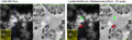
Hyperspectral Information of pre-segmented individual granule (LF).
Tutorial on Hyperspectral Analysis with 3D Slicer
Key Investigators
- Sungmin Hong (New York University)
- Guido Gerig (New York University)
Project Description
This project aims to offer a tool which makes use of 3D/4D ophthalmology data in different modalities to extract information which can compensate each other for richer analysis.
3D high resolution data (high resolution structured illumination microscopy, SIM) displays individual cells with sharp boundaries which are hard to be localized in 4D hyperspectral data (confocal multispectral laser scanning microscopy, LSM) because of low resolution.
The tool that we want to provide to users should offer image processing modules, such as, co-registration between SIM and LSM data, segmentation on SIM and mapping the segmentation label from SIM to LSM to analyze spectral information of each granule. The examination of spectral characteristics and statistics of granules, related with age and disease progress might reveal metabolism in human retina physiology.
| Objective
|
Approach and Plan
|
Progress and Next Steps
|
|
3D/4D Ophthalmology Image Anaylsis Framework
- Read 4D hyperspectral data
- Viewer and interactor for 3D hi-res image data and 4D hyperspectral data
- Co-registration between 3D hi-res data and 4D hyperspectral data
- Cell segmentation in 3D hi-res data
- Statistics of cells (possibly location, size, distribution)
- Plot of spectra of selected cells or a region-of-interest
|
- Review existing modules in 3D Slicer
- Review existing modules for 4D data viewer, such as, multi-volume viewer extension
- Try existing segmentation modules in Slicer to see if they can work on SIM data
- Review existing modules for cell statistics after segmentation
- Implementation/Integration
- Implement/integrate 4D hyperspectral data viewer to show image and spectral information.
- Integrate registration functionality for co-registration between 3D hi-res data and 4D hyperspectral data at image level
- Integrate user-initialized level set segmentation for cell segmentation or EM segmentation module
- Implement a viewer and an interactor for cell statistics
- User Manual
- Create an user manual to comprehend a overview of an extension
- Guide users to different extensions in a algorithmic flow chart if there are any desired functions (registration, segmentation, or etc.) which are already implemented in existing modules.
|
- Hyperspectral Analysis
- Implemented a module to convert 4D LSM data to a series of 3D data compatible to MultiVolume Explorer
- With a converted series of 3D data, MultiVolume explorer offered a basic analysis tool for hyperspectral data.
- Label statistics or segmentation need to be added in the future
- Registration
- Basic registration algorithms in Slicer worked well on linear registration between 3D SIM and a cropped and dimension reduced 4D LSM data.
- Detecting corresponding regions of 3D SIM in 4D LSM data needs to be developed in the future.
- Segmentation
- Watershed segmentation on 3D hi-res image data (WASP) was not successful.
- Editor/Segmentation Editor worked good on slice-by-slice segmentation
- Will investigate more on 3D segmentation capability of Slicer with possible collaboration with other groups.
|
Background and References
Acknowledgement
We would like to thank Dr. Thomas Ach (University Hospital Würzburg, Germany) for the significant contribution on the data and the project.