File list
From NAMIC Wiki
This special page shows all uploaded files.
| Date | Name | Thumbnail | Size | User | Description | Versions |
|---|---|---|---|---|---|---|
| 16:12, 14 May 2010 | Stochastic-i.png (file) |  |
23 KB | Marianna | 2 | |
| 16:07, 14 May 2010 | Lupus-i.png (file) |  |
15 KB | Marianna | 2 | |
| 16:03, 14 May 2010 | NAMIC-Kit-Overview-i.png (file) |  |
9 KB | Marianna | 1 | |
| 16:03, 14 May 2010 | Slicer3 logo-i.jpg (file) |  |
17 KB | Marianna | 1 | |
| 15:46, 14 May 2010 | Perkstation-i.jpg (file) |  |
7 KB | Marianna | 1 | |
| 15:29, 14 May 2010 | Child-i.png (file) |  |
17 KB | Marianna | 1 | |
| 15:15, 14 May 2010 | Schiz-thumb.png (file) |  |
13 KB | Marianna | 1 | |
| 15:11, 14 May 2010 | Schiz thumb.jpg (file) |  |
13 KB | Marianna | 4 | |
| 20:40, 13 May 2010 | Trptutorial thumb.jpg (file) |  |
160 KB | Kikinis | 2 | |
| 18:01, 13 May 2010 | BrainVentriclesAndLesions.png (file) |  |
182 KB | Mscully | Slice of brain overlaid with ventricles and lesions as volumes | 1 |
| 17:47, 13 May 2010 | CompareViewLDiffZoomedIn.jpg (file) |  |
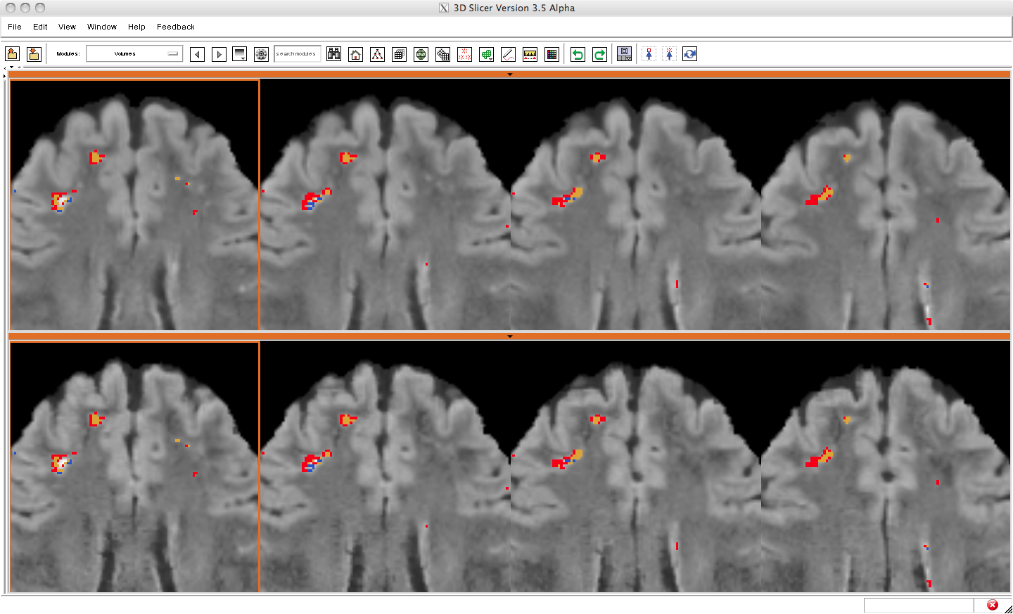
454 KB | Mscully | Compare Layout zoomed in with baseline and follow-up FLAIR images overlaid with the lesion difference image where yellow represents lesion in time1 and time2, red is lesion gained in time2, and blue is lesion lost in time2. | 1 |
| 17:43, 13 May 2010 | ComapareViewL1L2Overview.jpg (file) |  |
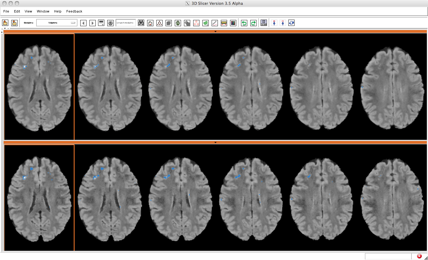
473 KB | Mscully | Compare Layout overview of difference between baseline and followup of lupus lesion subject with independent lesion segmentations | 1 |
| 17:21, 13 May 2010 | Case01045 2T tc.png (file) |  |
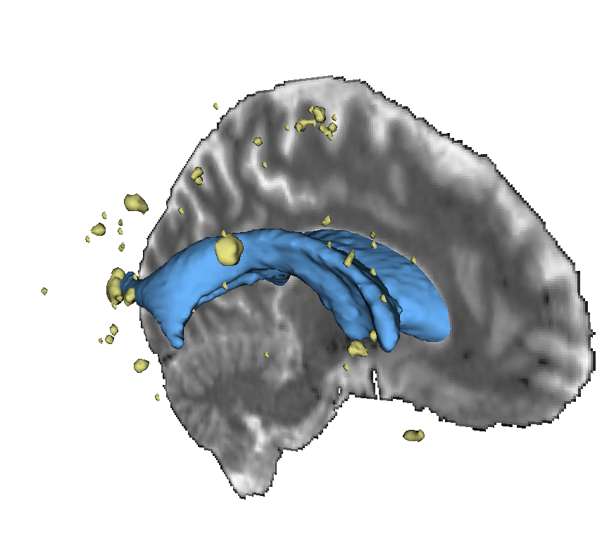
795 KB | Yrathi | Two tensor tractography starting from a seed region in the mid-sagittal plane of the corpus callosum. As is clear, the method is able to trace lateral transcallosal fibers through regions of crossing. | 1 |
| 15:41, 13 May 2010 | COPDGeneDashboard4.png (file) |  |
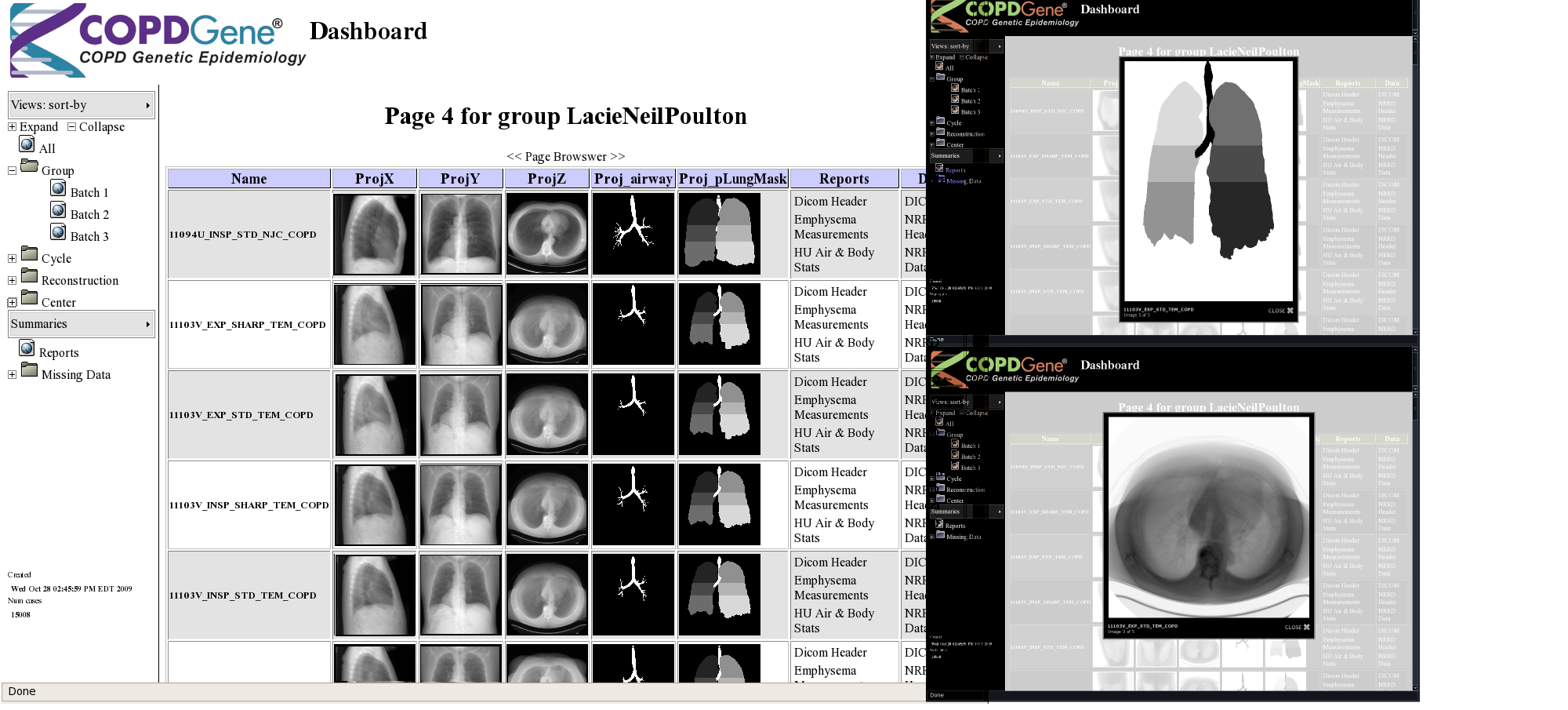
530 KB | Melonakos | 2 | |
| 14:20, 13 May 2010 | SlicerRegistrationLibrary-i.png (file) |  |
21 KB | Marianna | 1 | |
| 14:15, 13 May 2010 | CenterLine-i.png (file) |  |
7 KB | Marianna | 1 | |
| 14:15, 13 May 2010 | Primate-i.jpg (file) |  |
18 KB | Marianna | 1 | |
| 14:14, 13 May 2010 | Confocal-i.jpg (file) |  |
15 KB | Marianna | 1 | |
| 14:14, 13 May 2010 | Arctic-i.png (file) |  |
20 KB | Marianna | 1 | |
| 14:14, 13 May 2010 | TransRectal-i.png (file) |  |
17 KB | Marianna | 1 | |
| 14:14, 13 May 2010 | WhiteMatterLesions-i.png (file) |  |
14 KB | Marianna | 1 | |
| 21:00, 11 May 2010 | MoriAtlas Label+ColorLUTs.zip (file) |  |
50 KB | Meier | 1 | |
| 16:12, 11 May 2010 | ABC-MIND3.png (file) |  |
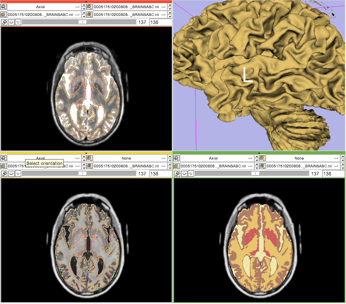
118 KB | Gerig | 1 | |
| 16:12, 11 May 2010 | ABC-MIND2.png (file) |  |
122 KB | Gerig | Hans Johnson, IOWA Mind Institute, modified ABC method demonstration | 1 |
| 16:11, 11 May 2010 | ABC-MIND1.png (file) |  |
594 KB | Gerig | Hans Johnson, IOWA Mind Institute, modified ABC method demonstration | 1 |
| 14:52, 11 May 2010 | SegHippoAmy.png (file) |  |
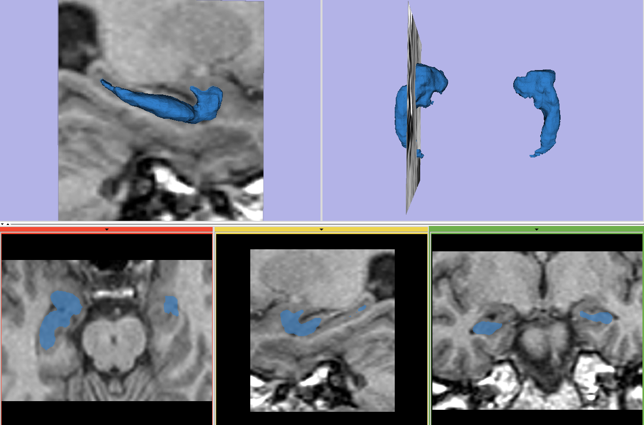
313 KB | Ygao | Segmentation of hippocampus and amygdala | 1 |
| 14:40, 11 May 2010 | RSSMandible.png (file) |  |
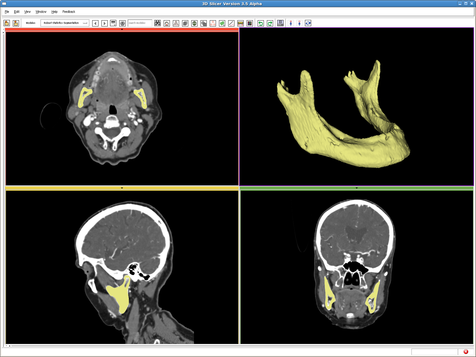
446 KB | Ygao | RSS segmentation of the mandible | 1 |
| 14:20, 11 May 2010 | TBI-seg.jpg (file) |  |
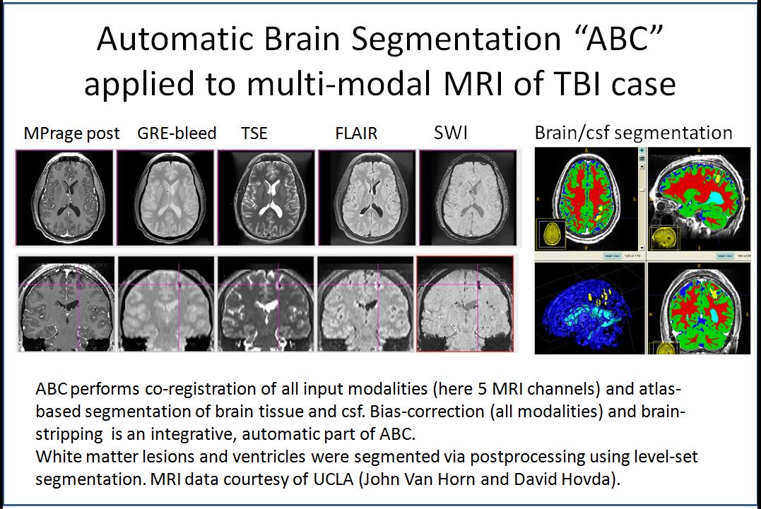
160 KB | Gerig | Automatic Brain Segmentation (ABC) applied to TBI multi-modality MRI. May 2010, Guido Gerig and Marcel Prastawa | 1 |
| 22:09, 10 May 2010 | Rview afib roi reg.gif (file) |  |
3.18 MB | Jfishbaugh | 1 | |
| 22:07, 10 May 2010 | Registerimages afib roi reg.gif (file) |  |
7.13 MB | Jfishbaugh | 1 | |
| 17:03, 10 May 2010 | Regtestbedmain.png (file) |  |
296 KB | Jfishbaugh | 1 | |
| 16:54, 10 May 2010 | Rview after.gif (file) |  |
1.75 MB | Jfishbaugh | 1 | |
| 16:53, 10 May 2010 | After.gif (file) |  |
2.2 MB | Jfishbaugh | 1 | |
| 16:53, 10 May 2010 | Before.gif (file) |  |
506 KB | Jfishbaugh | 1 | |
| 16:52, 10 May 2010 | Prevspost.png (file) |  |
562 KB | Jfishbaugh | 1 | |
| 16:51, 10 May 2010 | Rview tumor reg.gif (file) |  |
2.38 MB | Jfishbaugh | 1 | |
| 16:51, 10 May 2010 | Tumor before.gif (file) |  |
534 KB | Jfishbaugh | 1 | |
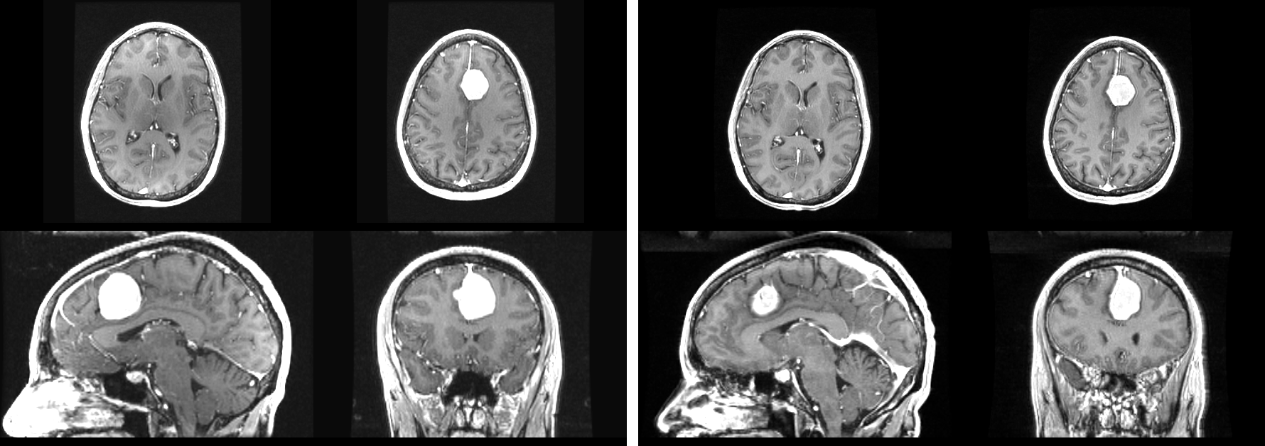
| 16:50, 10 May 2010 | All both.png (file) |  |
684 KB | Jfishbaugh | 1 | |
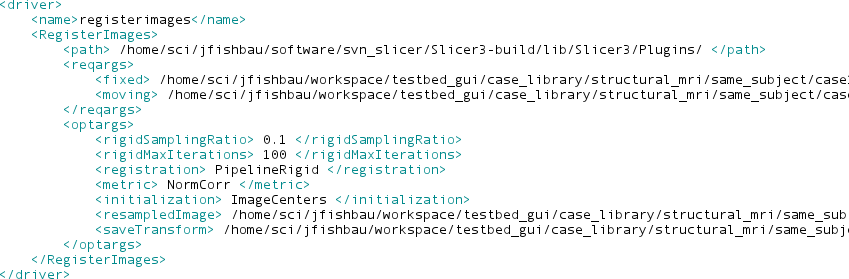
| 16:46, 10 May 2010 | Xmlexample.png (file) |  |
38 KB | Jfishbaugh | 1 | |
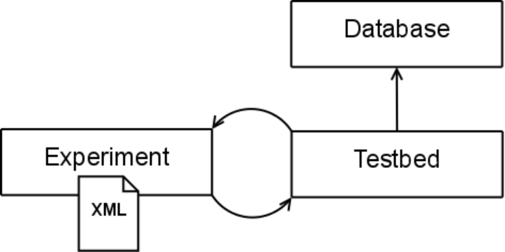
| 16:46, 10 May 2010 | Diagram1.png (file) |  |
26 KB | Jfishbaugh | 1 | |
| 22:58, 6 May 2010 | SPIE MI Gholami.pdf (file) |  |
193 KB | Ygao | @conference{gao2010segmentation, title={{Segmentation of the Endocardial Wall of the Left Atrium using Local Region-Based Active Contours and Statistical Shape Learning}}, author={Gao, Y. and Gholami, B. and MacLeod, R.S. and Blauer, J. and Haddad, W. | 1 |
| 00:59, 6 May 2010 | Collaboration ETH Come Image 1.png (file) |  |
45 KB | Tkapur | 1 | |
| 00:58, 6 May 2010 | Collaboration ETH Come Image 2.png (file) |  |
13 KB | Tkapur | 1 | |
| 00:29, 6 May 2010 | UtahSegGUI screen.png (file) |  |
11 KB | Prastawa | 1 | |
| 00:28, 6 May 2010 | UtahSegPlugin screen.png (file) |  |
21 KB | Prastawa | 2 | |
| 21:16, 4 May 2010 | Scully-FrontHumNeurosci2010-fig3.jpeg (file) |  |
153 KB | Marianna | 1 | |
| 15:18, 4 May 2010 | OCAIRO animation.gif (file) |  |
1,004 KB | Tkapur | 1 | |
| 12:48, 4 May 2010 | NA-MICSitePix.zip (file) | |
4.74 MB | Wjp | 1 | |
| 19:38, 2 May 2010 | Iowa-slicertraining.png (file) |  |
27 KB | Marianna | 1 | |
| 19:26, 1 May 2010 | N4itk lcov report detailed.jpg (file) |  |
127 KB | Fedorov | 1 |