File list
From NAMIC Wiki
This special page shows all uploaded files.
| Date | Name | Thumbnail | Size | User | Description | Versions |
|---|---|---|---|---|---|---|
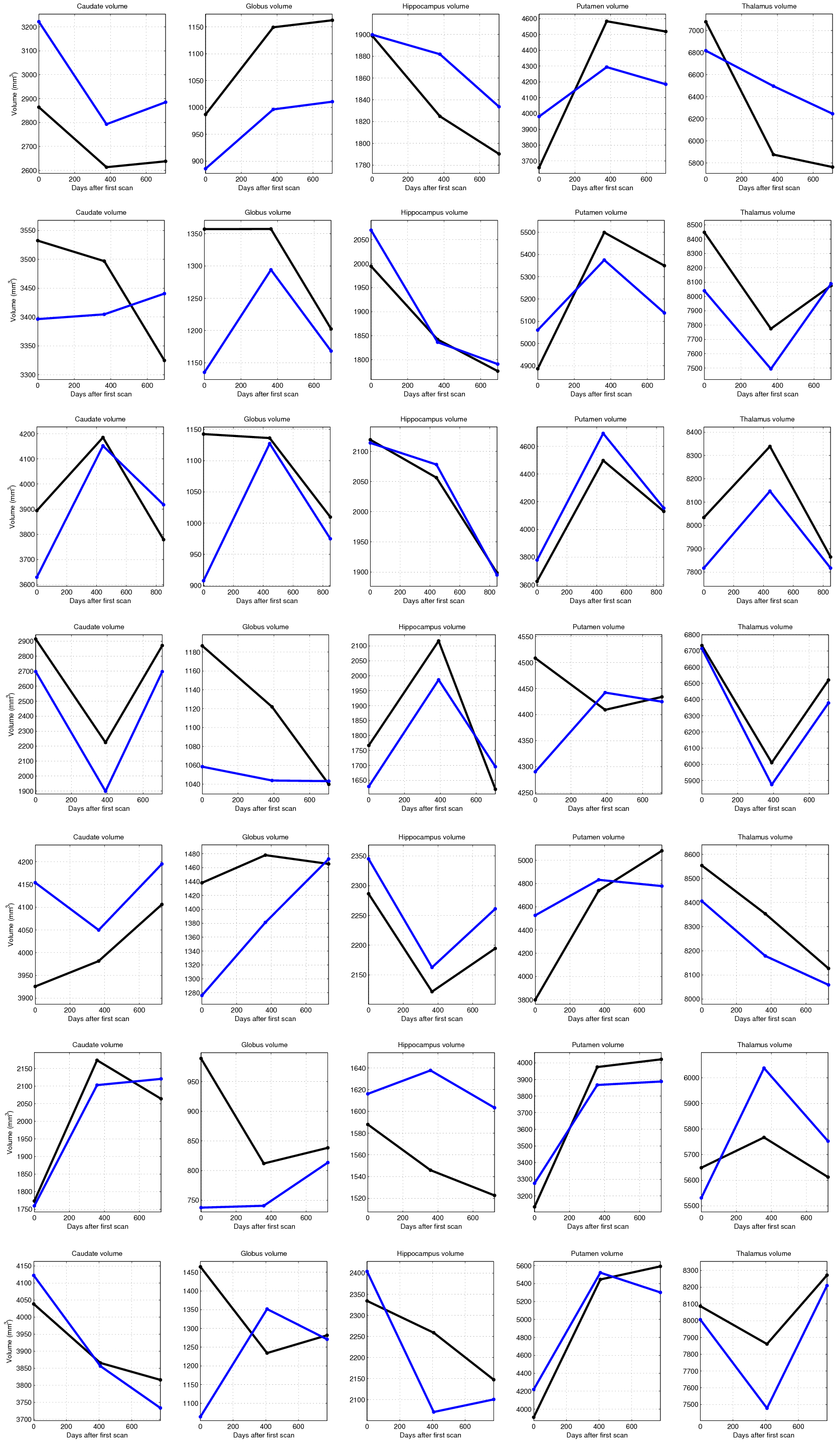
| 21:56, 11 January 2012 | CTRL all sub cort volume.png (file) |  |
406 KB | Jfishbaugh | 1 | |
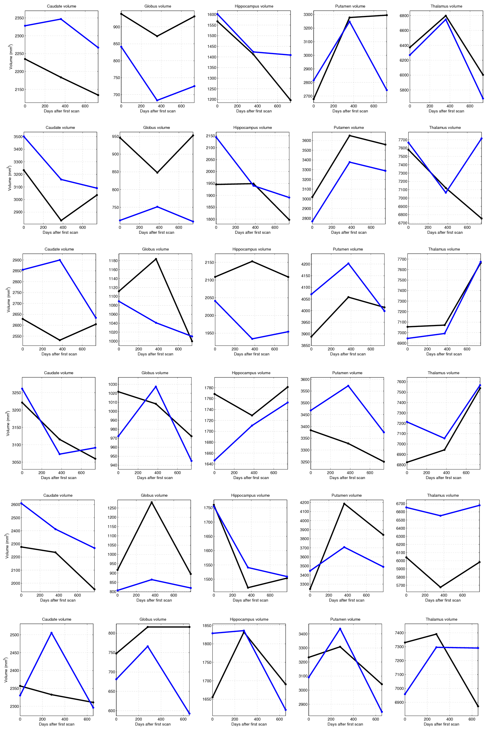
| 21:56, 11 January 2012 | HD all sub cort volume.png (file) |  |
396 KB | Jfishbaugh | 1 | |
| 18:00, 11 January 2012 | LiveUltrasound2012Jan.pdf (file) | 418 KB | Ungi | 2 | ||
| 16:24, 11 January 2012 | 2012-01-12-sharp.pdf (file) | 3.23 MB | Gregsharp | 1 | ||
| 15:18, 11 January 2012 | PlusIntro2012Jan.pdf (file) | 1.43 MB | Lasso | Introduction to the Plus (Public software library for ultrasound imaging research) toolkit | 1 | |
| 02:01, 11 January 2012 | DICOMRTImportInSlicer4.pptx (file) | 815 KB | Pinter | DICOM RT Import in Slicer 4 slides | 1 | |
| 22:56, 10 January 2012 | 2012-01-09-fedorov-qin slicer annotation.pdf (file) | 811 KB | Fedorov | 2 | ||
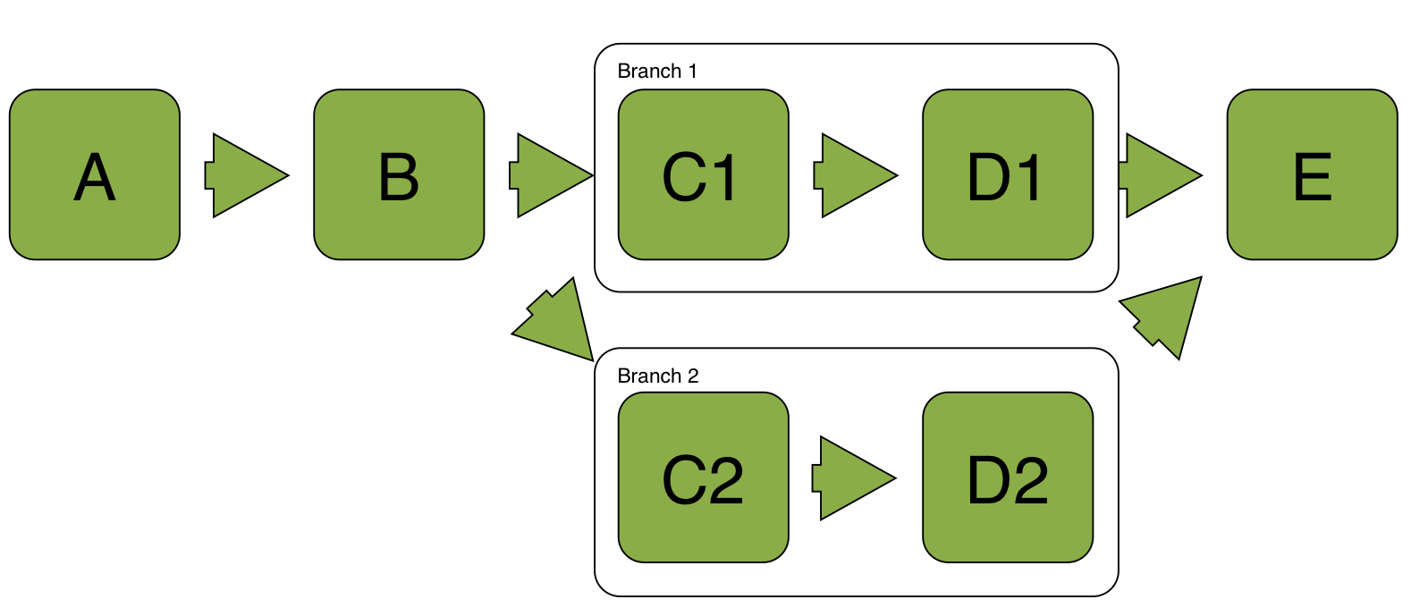
| 16:57, 10 January 2012 | Workflow scheme.jpg (file) |  |
73 KB | Demian | 1 | |
| 15:48, 10 January 2012 | 2012-01-10-Linking.png (file) |  |
44 KB | Kikinis | 1 | |
| 15:29, 10 January 2012 | DemoTuto2Slicer.mpeg (file) | 22.13 MB | JChris.FillionR | QtTesting Demo | 1 | |
| 15:16, 10 January 2012 | AHM-2012-SlicerTesting.pdf (file) | 139 KB | JChris.FillionR | QtTesting Presentation | 1 | |
| 15:01, 10 January 2012 | LoadableModules.pptx (file) | 6.16 MB | Finetjul | 1 | ||
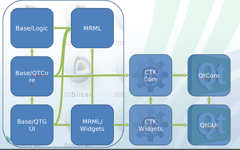
| 09:13, 10 January 2012 | AHM2012 SlicerArch.png (file) |  |
196 KB | JChris.FillionR | 2 | |
| 05:29, 10 January 2012 | 2012-01-09-sharp-dicomrt.pdf (file) | 127 KB | Gregsharp | 1 | ||
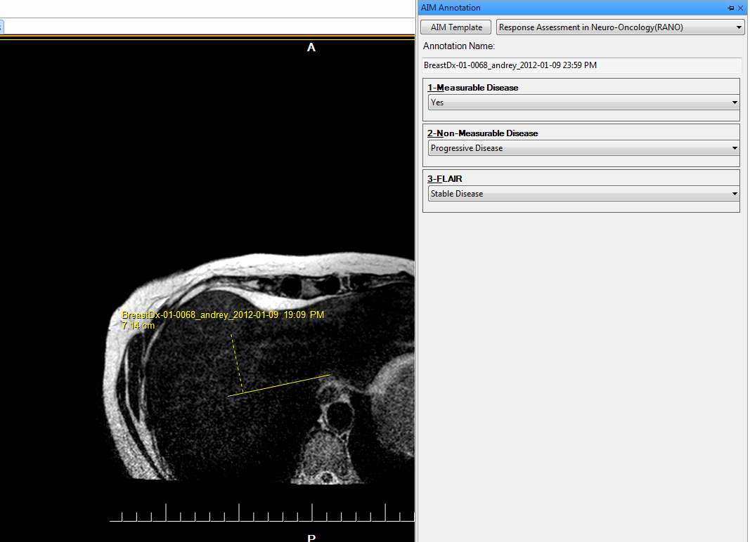
| 05:07, 10 January 2012 | AIM template demo.jpg (file) |  |
171 KB | Fedorov | 1 | |
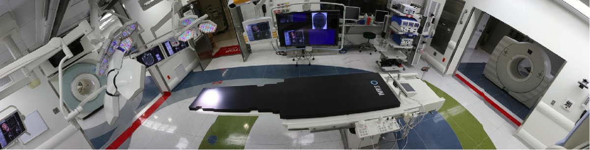
| 00:25, 10 January 2012 | 1200px-AMIGOJuly2011-1.png (file) | 520 KB | Dmwelch | AMIGO surgery suite | 1 | |
| 20:28, 9 January 2012 | Laser-ablation-image.png (file) |  |
62 KB | Inorton | 1 | |
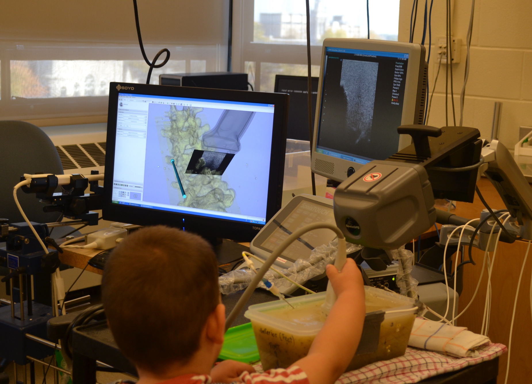
| 20:11, 9 January 2012 | SpineUS-Levi.JPG (file) |  |
774 KB | Ungi | 1 | |
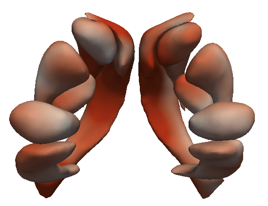
| 20:02, 9 January 2012 | HD sub cort shapes.png (file) |  |
136 KB | Jfishbaugh | 1 | |
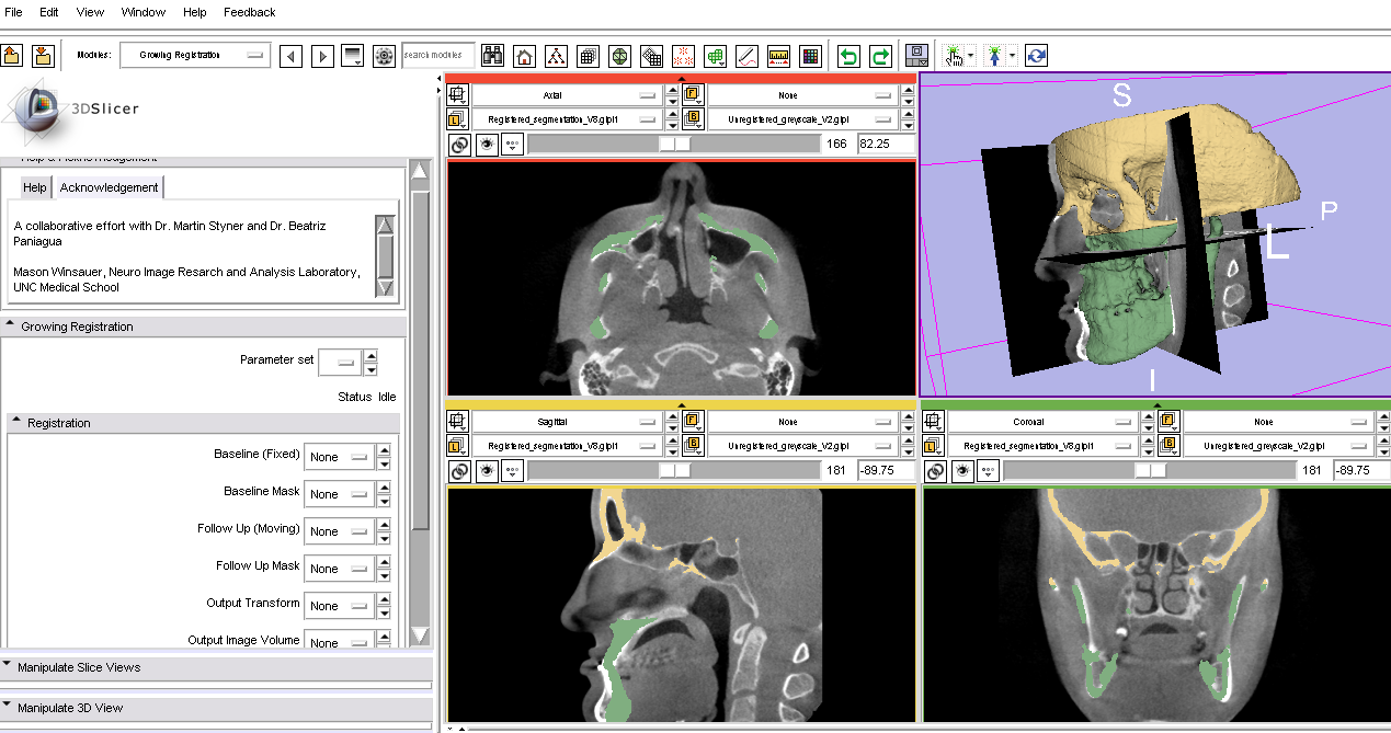
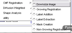
| 19:01, 9 January 2012 | CMFreg02.png (file) |  |
288 KB | Bpaniagua | 1 | |
| 19:01, 9 January 2012 | CMFreg01.png (file) |  |
13 KB | Bpaniagua | paniagua project week 2012 | 1 |
| 17:47, 9 January 2012 | Carma fid misreg.png (file) |  |
243 KB | Ggardner | 2 | |
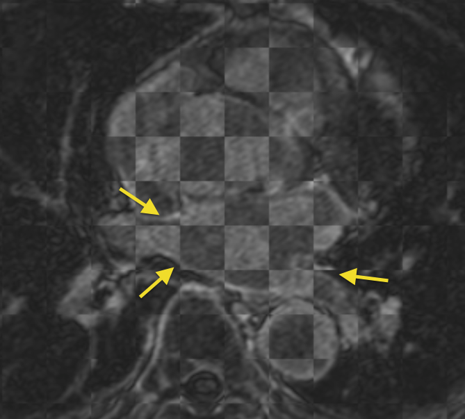
| 17:42, 9 January 2012 | Carma PV misreg.png (file) |  |
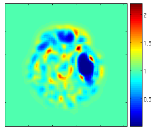
268 KB | Ggardner | An example of the misregistration that is seen when trying to register the pulmonary veins of pre- and post-ablation LGE-MRI scans from the same patient (scans acquired roughly 3 months apart). | 1 |
| 15:27, 9 January 2012 | Fig1.png (file) |  |
92 KB | Rtibrewal | 2 | |
| 15:25, 9 January 2012 | Fig2.png (file) |  |
90 KB | Rtibrewal | 1 | |
| 12:39, 9 January 2012 | RadnosticsWorkFlow.png (file) | 4 KB | AnthonyBlumfield | 1 | ||
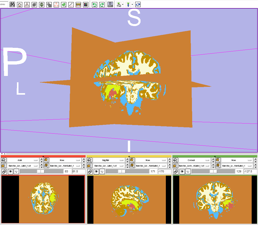
| 06:44, 9 January 2012 | TBI Seg lable Slicer.png (file) |  |
39 KB | Bowang | 1 | |
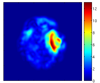
| 05:57, 9 January 2012 | DefVis VecMag TBISeg.png (file) |  |
15 KB | Bowang | 1 | |
| 05:54, 9 January 2012 | DefVis DetJacobian TBISeg.png (file) |  |
19 KB | Bowang | 2 | |
| 00:52, 9 January 2012 | Carma norm LAA.png (file) |  |
98 KB | Ggardner | Reverted to version as of 00:51, 9 January 2012 | 4 |
| 00:51, 9 January 2012 | Carma vert LAA.png (file) |  |
148 KB | Ggardner | 2 | |
| 00:30, 9 January 2012 | Carma no reflow.png (file) |  |
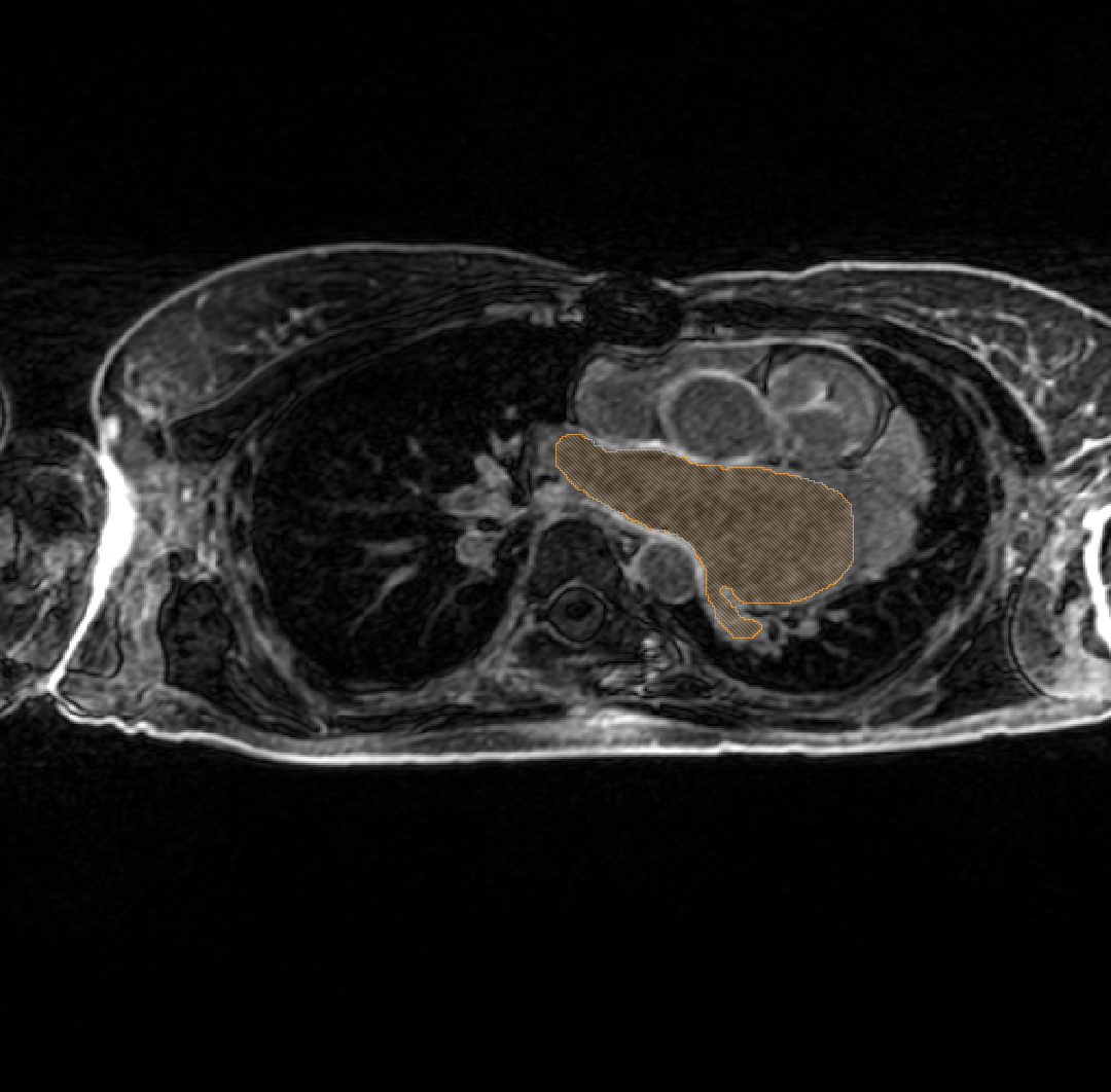
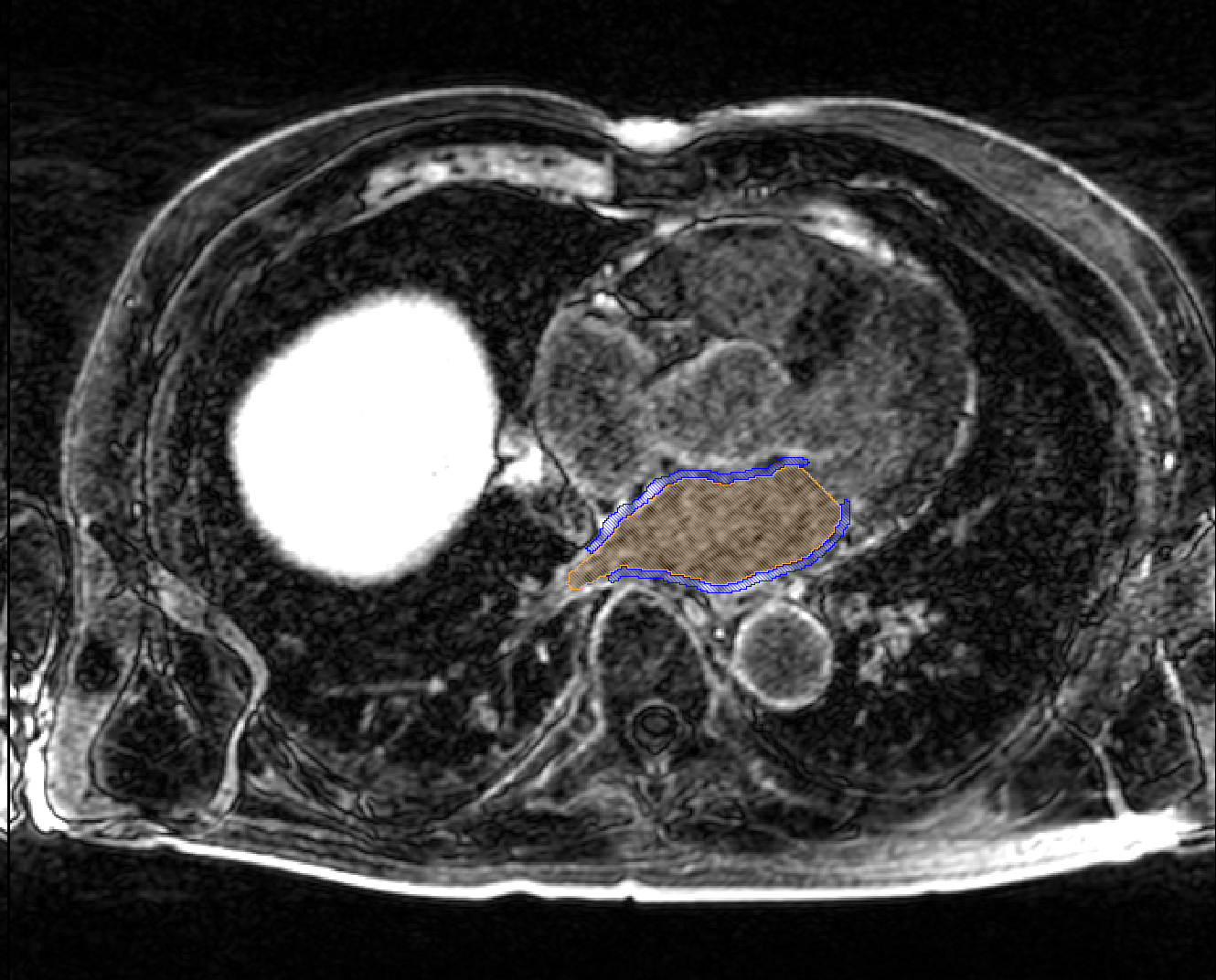
327 KB | Ggardner | LGE-MRI scans acquired immediately post-ablation pose extra challenges for registration; often times, edema and other acute physiological changes effect the washout kinetics of the contrast agent and the morphology of the LA. Here the MR contrast agent h | 1 |
| 00:24, 9 January 2012 | Carma hypertrophy.png (file) |  |
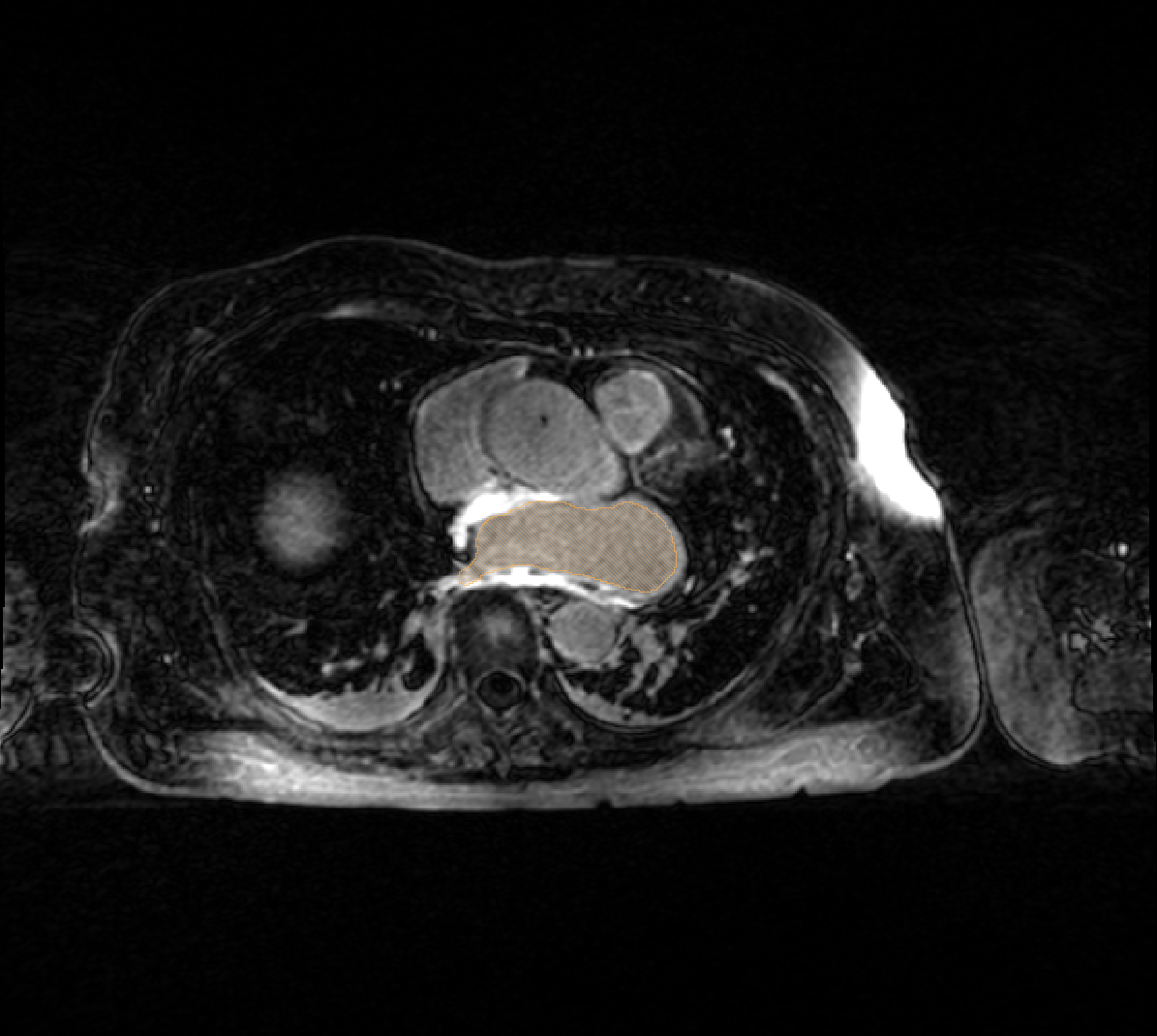
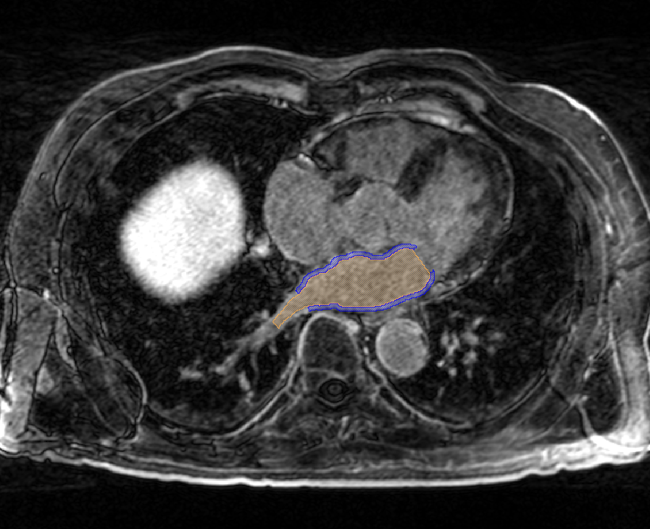
303 KB | Ggardner | An example of a hypertrophied LA, the extent of which is denoted by the orange label mask. | 1 |
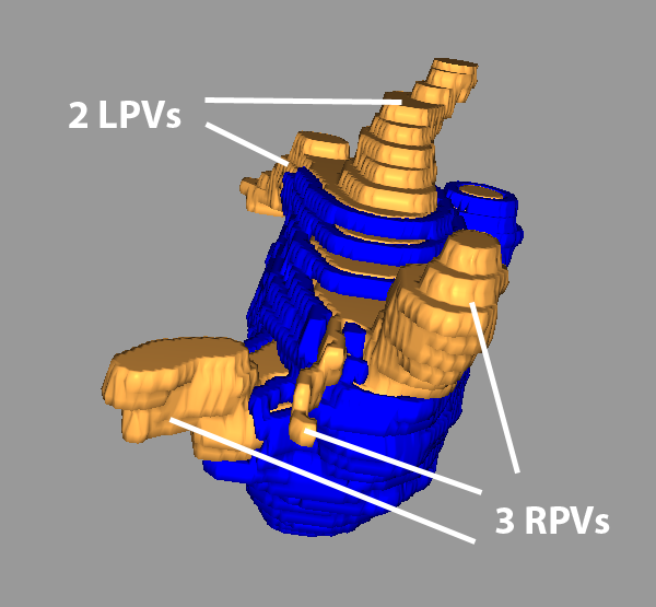
| 23:25, 8 January 2012 | Carma 3RPV.png (file) |  |
148 KB | Ggardner | A LA with the 2 left PVs (typical) and an abnormal 3 right PVs. | 1 |
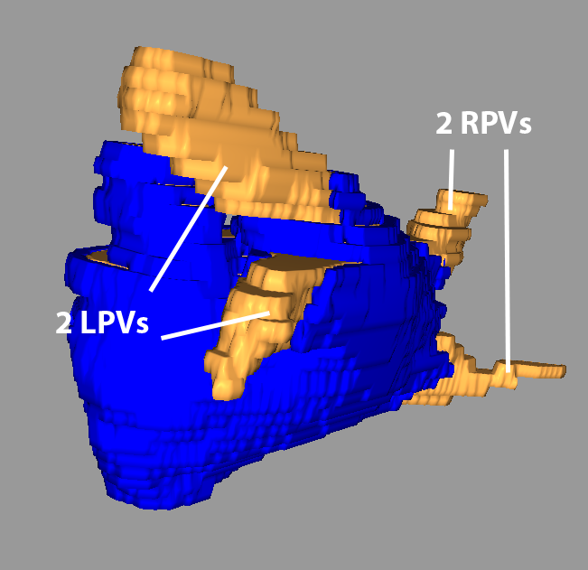
| 23:22, 8 January 2012 | Carma 4RPV.png (file) |  |
193 KB | Ggardner | A LA with the 2 left PVs (typical) and an abnormal 4 right PVs. | 1 |
| 23:03, 8 January 2012 | Carma 3RPVs.png (file) |  |
148 KB | Ggardner | Reverted to version as of 00:13, 7 January 2012 | 4 |
| 23:39, 7 January 2012 | Slicer4 CLI.ppt (file) | 749 KB | Millerjv | 2 | ||
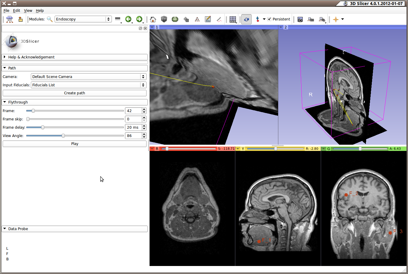
| 22:53, 7 January 2012 | 3D Slicer 4.0.1.2012-01-07 175.png (file) |  |
373 KB | Pieper | 1 | |
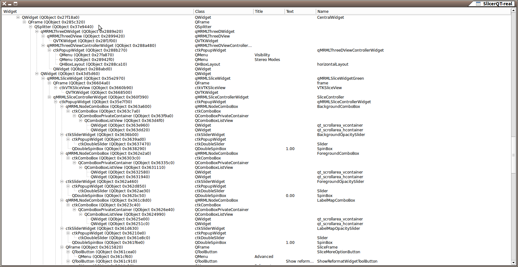
| 22:47, 7 January 2012 | SlicerQT-real 174.png (file) |  |
221 KB | Pieper | 1 | |
| 00:00, 7 January 2012 | Carma Norm LPVs.png (file) |  |
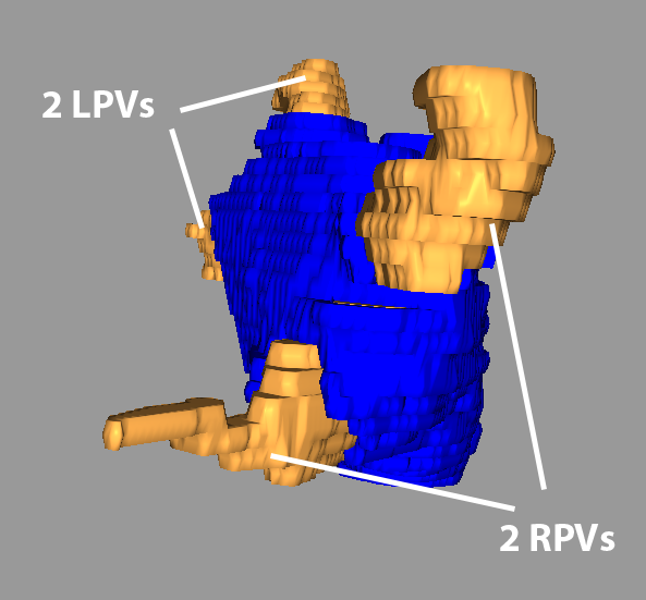
138 KB | Ggardner | A typical arrangement of the PVs: 2 distinct LPVs and 2 RPVs. | 1 |
| 00:00, 7 January 2012 | Carma Norm RPVs.png (file) |  |
115 KB | Ggardner | A typical arrangement of the PVs: 2 distinct LPVs and 2 RPVs. | 1 |
| 23:49, 6 January 2012 | Carma RPVs small.png (file) |  |
130 KB | Ggardner | Variations in the size of the PVs also occurs; here the inferior PV is dramatically smaller than normal and the superior larger than is typical. | 1 |
| 23:47, 6 January 2012 | Carma Com LPV.png (file) |  |
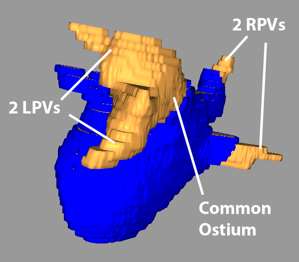
122 KB | Ggardner | A LA with the typical number of PVs (2 left, 2 right); however, the 2 left PVs share a common trunk. | 1 |
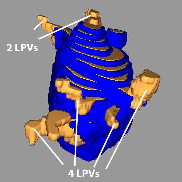
| 23:46, 6 January 2012 | Carma 4rpvs.png (file) |  |
165 KB | Ggardner | A LA with the 2 left PVs (typical) and an abnormal 4 right PVs. | 1 |
| 22:44, 6 January 2012 | Carma ex pre.png (file) |  |
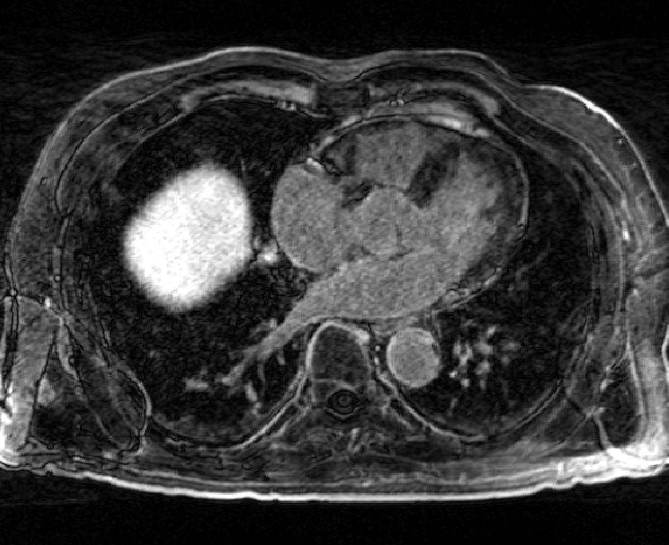
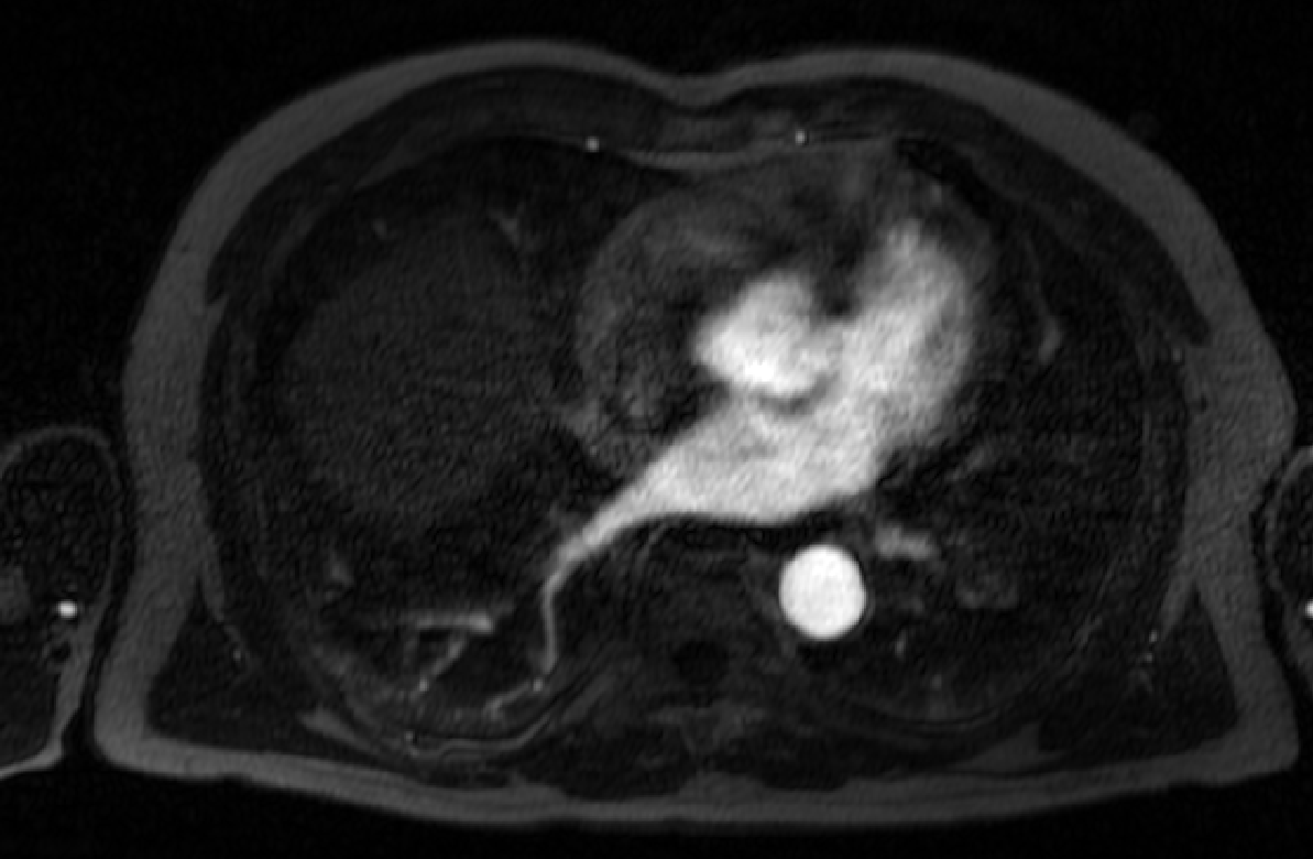
267 KB | Ggardner | Carma Center example pre-ablation LGE-MRI image | 1 |
| 22:43, 6 January 2012 | Carma ex pre seg.png (file) |  |
388 KB | Ggardner | Carma Center example pre-ablation LGE-MRI image, with manually segmented LA wall and blood pool overlaid | 1 |
| 22:43, 6 January 2012 | Carma ex post.png (file) |  |
276 KB | Ggardner | Carma Center example post-ablation LGE-MRI image | 1 |
| 22:42, 6 January 2012 | Carma ex post seg.png (file) |  |
393 KB | Ggardner | Carma Center example post-ablation LGE-MRI image, with manual segmentations of the LA wall and blood pool overlaid | 1 |
| 22:41, 6 January 2012 | Carma ex mra.png (file) |  |
179 KB | Ggardner | Carma Center example MRA image (acquired prior to LGE-MRI image) | 1 |
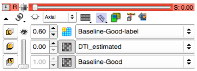
| 16:47, 6 January 2012 | 2012-01-FullViewerController.png (file) |  |
32 KB | Kikinis | 3 |