Difference between revisions of "NA-MIC Internal Collaborations:DiffusionImageAnalysis"
| (12 intermediate revisions by the same user not shown) | |||
| Line 16: | Line 16: | ||
Age-associated alterations in cortical gray and white matter signal intensity and gray to white matter contrast. Neuroimage, 2009 Oct 15;48(1):21-8 | Age-associated alterations in cortical gray and white matter signal intensity and gray to white matter contrast. Neuroimage, 2009 Oct 15;48(1):21-8 | ||
| − | |||
|- | |- | ||
| − | | | [[Image: | + | | | [[Image:GTTubSurfaceSeg-Img1.png|200px]] |
| | | | | | ||
| − | == [[Projects: | + | == [[Projects:TubularSurfaceSegmentation|Tubular Surface Segmentation Framework]] == |
| − | |||
| − | |||
| − | | | + | We have proposed a new model for tubular surfaces that transforms the problem of detecting a surface in 3D space, to detecting a curve in 4D space. Besides allowing us to impose a "soft" tubular shape prior, this also leads to computational efficiency over conventional surface segmentation approaches. [[Projects:TubularSurfaceSegmentation|More...]] |
| − | + | <font color="red">'''New: '''</font> V. Mohan, G. Sundaramoorthi, M. Kubicki and A. Tannenbaum. Tubular Surface Evolution for Segmentation of the Cingulum Bundle From DW-MRI. September 2008. Proceedings of the Second Workshop on Mathematical Foundations of Computational Anatomy (MFCA'08), Int Conf Med Image Comput Comput Assist Interv. 2008. | |
| − | |||
| − | = | + | <font color="red">'''New: '''</font> V. Mohan, G. Sundaramoorthi and A. Tannenbaum. Tubular Surface Segmentation for Extracting Anatomical Structures from Medical Imagery (in submission). IEEE Transactions on Medical Imaging. |
| − | |||
| − | |||
| − | |||
| − | |||
| − | |||
| − | |||
| − | |||
| − | |||
| − | |||
| − | |||
| − | |||
| − | |||
| − | |||
| − | |||
| − | |||
| − | |||
|- | |- | ||
| − | | | [[Image: | + | | | [[Image:GT-PopStudyVis OnCBs Case19-View2.jpg|200px]] |
| | | | | | ||
| − | == [[Projects: | + | == [[Projects:TubularSurfaceSegmentationPopStudy|Group Study on DW-MRI using the Tubular Surface Model]] == |
| − | |||
| − | |||
| − | + | We have proposed a new framework for performing group studies on DW-MRI data sets using the Tubular Surface Model of Mohan et al. We successfully apply this framework to discriminating schizophrenic cases from normal controls, as well as towards visualizing the regions of the Cingulum Bundle that are affected by Schizophrenia. [[Projects:TubularSurfaceSegmentationPopStudy|More...]] | |
| − | |||
| − | |||
| − | |||
| − | |||
| − | = | + | <font color="red">'''New: '''</font> V. Mohan, G. Sundaramoorthi, M. Kubicki and A. Tannenbaum. Population Analysis of the Cingulum Bundle using the Tubular Surface Model for Schizophrenia Detection. SPIE Medical Imaging 2010 (in submission). |
| − | |||
|- | |- | ||
| + | | | [[Image:ZoomedResultWithModel.png|200px]] | ||
| + | | | | ||
| + | == [[Projects:GeodesicTractographySegmentation|Geodesic Tractography Segmentation]] == | ||
| + | In this work, we provide an energy minimization framework which allows one to find fiber tracts and volumetric fiber bundles in brain diffusion-weighted MRI (DW-MRI). [[Projects:GeodesicTractographySegmentation|More...]] | ||
| − | + | <font color="red">'''New: '''</font> J. Melonakos, E. Pichon, S. Angenet, and A. Tannenbaum. Finsler Active Contours. IEEE Transactions on Pattern Analysis and Machine Intelligence, March 2008, Vol 30, Num 3. | |
| − | |||
| − | |||
| − | |||
| − | |||
| − | |||
|} | |} | ||
| − | === | + | === Clustering and Quantitative Analysis === |
{| cellpadding="10" style="text-align:left;" | {| cellpadding="10" style="text-align:left;" | ||
| − | | style="width:15%" | [[Image: | + | | style="width:15%" | [[Image:Cbg-dtiatlas-tracts.png|200px]] |
| style="width:85%" | | | style="width:85%" | | ||
| − | |||
| − | + | == [[Projects:DTIPopulationAnalysis|Group Analysis of DTI Fiber Tracts]] == | |
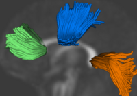
| − | | | + | Analysis of populations of diffusion images typically requires time-consuming manual segmentation of structures of interest to obtain correspondance for statistics. This project uses non-rigid registration of DTI images to produce a common coordinate system for hypothesis testing of diffusion properties. [[Projects:DTIPopulationAnalysis|More...]] |
| − | + | <font color="red">'''New: '''</font> Casey B. Goodlett, P. Thomas Fletcher, John H. Gilmore, Guido Gerig. Group Analysis of DTI Fiber Tract Statistics with Application to Neurodevelopment. NeuroImage 45 (1) Supp. 1, 2009. p. S133-S142. | |
| − | |||
| − | |||
| − | |||
| − | |||
| − | |||
|- | |- | ||
| − | |||
| − | |||
| − | |||
| − | |||
| − | |||
| − | |||
| − | |||
| − | |||
| − | |||
| − | |||
| − | |||
| − | |||
| − | |||
| − | |||
| − | |||
| − | |||
| − | |||
|} | |} | ||
| Line 138: | Line 87: | ||
|- | |- | ||
| − | |||
| − | |||
| − | |||
| − | |||
| − | |||
| − | |||
| − | |||
|} | |} | ||
Latest revision as of 18:38, 4 November 2009
Home < NA-MIC Internal Collaborations:DiffusionImageAnalysisBack to NA-MIC Internal Collaborations
Diffusion Image Analysis
Tractography Methods
|
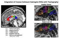
Corpus Callosum Fiber TractographyThe goal of this project is to examine the integrity of fibers in the corpus callosum in patients with schizophrenia and determine whether this is associated with brain activation during memory tasks. More... New: Salat D.H., Lee S.Y., van der Kouwe A.J., Greve D.N., Fischl B., Rosas H.D. Age-associated alterations in cortical gray and white matter signal intensity and gray to white matter contrast. Neuroimage, 2009 Oct 15;48(1):21-8 |
|
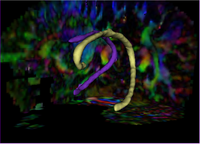
Tubular Surface Segmentation FrameworkWe have proposed a new model for tubular surfaces that transforms the problem of detecting a surface in 3D space, to detecting a curve in 4D space. Besides allowing us to impose a "soft" tubular shape prior, this also leads to computational efficiency over conventional surface segmentation approaches. More... New: V. Mohan, G. Sundaramoorthi, M. Kubicki and A. Tannenbaum. Tubular Surface Evolution for Segmentation of the Cingulum Bundle From DW-MRI. September 2008. Proceedings of the Second Workshop on Mathematical Foundations of Computational Anatomy (MFCA'08), Int Conf Med Image Comput Comput Assist Interv. 2008. New: V. Mohan, G. Sundaramoorthi and A. Tannenbaum. Tubular Surface Segmentation for Extracting Anatomical Structures from Medical Imagery (in submission). IEEE Transactions on Medical Imaging.
|
|
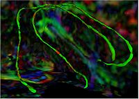
Group Study on DW-MRI using the Tubular Surface ModelWe have proposed a new framework for performing group studies on DW-MRI data sets using the Tubular Surface Model of Mohan et al. We successfully apply this framework to discriminating schizophrenic cases from normal controls, as well as towards visualizing the regions of the Cingulum Bundle that are affected by Schizophrenia. More...
|
|
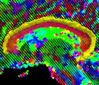
Geodesic Tractography SegmentationIn this work, we provide an energy minimization framework which allows one to find fiber tracts and volumetric fiber bundles in brain diffusion-weighted MRI (DW-MRI). More... New: J. Melonakos, E. Pichon, S. Angenet, and A. Tannenbaum. Finsler Active Contours. IEEE Transactions on Pattern Analysis and Machine Intelligence, March 2008, Vol 30, Num 3. |
Clustering and Quantitative Analysis
|
Group Analysis of DTI Fiber TractsAnalysis of populations of diffusion images typically requires time-consuming manual segmentation of structures of interest to obtain correspondance for statistics. This project uses non-rigid registration of DTI images to produce a common coordinate system for hypothesis testing of diffusion properties. More... New: Casey B. Goodlett, P. Thomas Fletcher, John H. Gilmore, Guido Gerig. Group Analysis of DTI Fiber Tract Statistics with Application to Neurodevelopment. NeuroImage 45 (1) Supp. 1, 2009. p. S133-S142. |
Validation
|
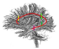
Contrasting Tractography MeasuresThis project represents a new initiative to build upon a shared vision among Cores 1, 3 and 5 that the field of medical image analysis would be well served by work in the area of validation, calibration and assessment of reliability in DW-MRI image analysis. More... New: S. Pujol, C-F. Westin, R. Whitaker, G. Gerig, T. Fletcher, V. Magnotta, S. Bouix, R. Kikinis, W. M. Wells III, and R. Gollub. Preliminary Results on the use of STAPLE for evaluating DT-MRI tractography in the absence of ground truth. In Proceedings of the 17th Annual Meeting of the International Society for Magnetic Resonance in Medicine ISMRM 2009, April 18-24, 2009. Honolulu, Hawaii. ISMRM 2009
|