Difference between revisions of "DBP2:MIND:Roadmap"
From NAMIC Wiki
m (→Roadmap: Centered images) |
|||
| (11 intermediate revisions by 2 users not shown) | |||
| Line 3: | Line 3: | ||
=Brain Lesion Analysis in Neuropsychiatric Systemic Lupus Erythematosus= | =Brain Lesion Analysis in Neuropsychiatric Systemic Lupus Erythematosus= | ||
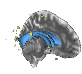
| + | [[Image:BrainVentriclesAndLesions.png|thumb|280px|Lesion volumes in yellow with ventricles and brain slice]] | ||
=='''Objective'''== | =='''Objective'''== | ||
This page describes the technology roadmap for lesion analysis in the NA-MIC Kit. Our objective is to create an end-to-end application allowing individual analysis of white matter lesions. This workflow applied to lupus patients is one of goals of the [[DBP2:MIND:Introduction|MIND DBP]]. The basic components necessary for this project are: | This page describes the technology roadmap for lesion analysis in the NA-MIC Kit. Our objective is to create an end-to-end application allowing individual analysis of white matter lesions. This workflow applied to lupus patients is one of goals of the [[DBP2:MIND:Introduction|MIND DBP]]. The basic components necessary for this project are: | ||
| Line 18: | Line 19: | ||
<center> | <center> | ||
{| | {| | ||
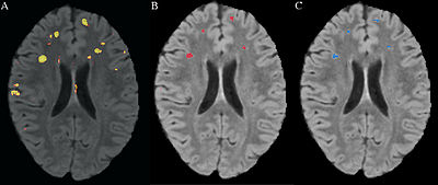
| − | |[[Image:Scully Figure3.jpg|thumb|400px| | + | |[[Image:Scully Figure3.jpg|thumb|400px|A. FLAIR with predicted probability of being lesion, B. FLAIR with manual segmentation, C. FLAIR with predicted probability after thresholding]] |
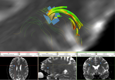
| − | |[[Image:TractsLesionZoomOverlay.png|thumb|380px|White Matter Lesion | + | |[[Image:TractsLesionZoomOverlay.png|thumb|380px|White Matter Lesion Disruption of a U fiber]] |
| + | |} | ||
| + | {| | ||
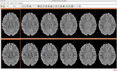
| + | |[[Image:ComapareViewL1L2Overview.jpg|thumb|400px| Compare Layout overview with baseline FLAIR image on top and followup FLAIR on bottom. Each image is overlaid with that time point's predicted lesion segmentation.]] | ||
| + | |[[Image:CompareViewLDiffZoomedIn.jpg|thumb|400px| Compare Layout zoomed in. Baseline FLAIR on top and followup on bottom. Images overlaid with a label map that represents changes in lesion. Yellow represents lesion that's unchanged between baseline and followup, red represents new lesion in the followup, and blue represents lesion present at baseline that is no longer present at followup. ]] | ||
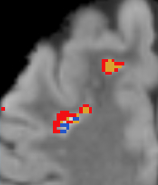
| + | | [[Image:MIND-DBPLesionClassZoomDetail.png|thumb|158px| Zoomed detail. ]] | ||
|} | |} | ||
</center> | </center> | ||
| Line 25: | Line 31: | ||
===Software=== | ===Software=== | ||
*We have a very active project for this pipeline on the NITRC resource [http://www.nitrc.org/frs/?group_id=180 3DSlicerLupusLesionModule] | *We have a very active project for this pipeline on the NITRC resource [http://www.nitrc.org/frs/?group_id=180 3DSlicerLupusLesionModule] | ||
| − | + | *The module can be checked out in Slicer3.6 using the extension manager or downloaded [http://www.nitrc.org/frs/download.php/570/LesionSegmentationApplications.tgz here]. | |
| − | + | *Source code is available via [http://www.nitrc.org/plugins/scmcvs/cvsweb.php/lupuslesion/LesionSegmentationApplications/?cvsroot=lupuslesion cvs]. | |
=== Tutorial === | === Tutorial === | ||
| Line 32: | Line 38: | ||
* The data needed to run the tutorial is available [http://www.nitrc.org/frs/download.php/569/LesionSegmentationTutorialData.tgz here]. | * The data needed to run the tutorial is available [http://www.nitrc.org/frs/download.php/569/LesionSegmentationTutorialData.tgz here]. | ||
| − | === | + | === Schedule === |
| + | * 06/21/2010 - 06/25/2010 Release new version of module and tutorial including directions and data for longitudinal analysis at the Summer project week | ||
| + | * 04/20/2010 Began updating module to use ITK review statistics | ||
| + | * 03/19/2010 Segmentation Method paper published in Frontiers in Human Neuroscience | ||
| + | * 01/04/2010 - 01/08/2010 Demonstrated longitudinal analysis and some of the multiscale work at the Winter Project Week / All Hands Meeting | ||
| + | * 12/15/2009 Developed longitudinal analysis method | ||
| + | * 11/18/2009 Submitted paper on novel lesion segmentation method to Frontiers in Human Neuroscience | ||
| + | * 08/15/2009 Expanded lesion model to include additional cases | ||
| + | * 06/22/2009 - 06/26/2009 Summer Project Week | ||
| + | * 06/18/2009 - 06/23/2009 Attended Human Brain Mapping Conference with submitted abstract on lesion classification using novel method | ||
| + | * 03/25/2009 - 04/02/2009 AAN conference outreach activity | ||
| + | * 03/01/2009 Released final version of the novel lesion segmentation method as a Slicer module | ||
| + | * 01/11/2009 Submitted abstract on novel lesion method for HBM 2009 | ||
| + | * 01/06/2009 Initial version of the novel lesion segmentation method released as slicer module. | ||
| + | * 01/05/2009 - 01/09/2009 Winter Project Week / All Hands Meeting | ||
| + | * 11/15/2008 - 11/19/2008 Abstract on initial version of the novel lesion method submitted to Society For Neuroscience conference and held a Conference / Outreach activity on the Slicer lesion segmentation tool. | ||
| + | * 09/11/2008 Began development on novel lesion segmentation method | ||
| + | * 09/06/2008 - 09/10/2008 Participated in the MS lesion segmentation challenge, part of the 3D Segmentation in the Clinic: A Grand Challenge II | ||
| + | * 08/14/2008 EMSegment has been run against collected lupus cases. Segmentations have improved but are still unusable | ||
| + | * 07/01/2008 Made tutorial and sample data available to the scientific community | ||
| + | * 06/23/2008 - 06/27/2008 Presented lesion segmentation tutorial at the Summer Project Week | ||
| + | * 05/12/2008 Submitted SFN abstract on a lesion segmentation method based on Vince Magnotta's work | ||
| + | * 04/01/2008 Gasparovic has manually traced all tutorial cases with lesions using T2/Flair images following tracing guidelines and a qualitative neuroradiological review report | ||
| + | * 03/31/2008 Completed collection of 5 lupus subjects on clinical sequence and optimized 3T sequence | ||
| + | * 03/17/2008 Applied itkEMS to sample lesion cases, results are shown [[DBP2:MIND:itkEMSResults|here]] | ||
| + | * 03/15/2008 Applied Vince Magnotta's lesions segmentation method to sample lesion cases, results are shown [[DBP2:MIND:itkBayesianLesion|here]] | ||
| + | * 02/05/2008 Created intensity standardization method | ||
| + | * 01/10/2008 Performed initial segmentations using EMSegment and submitted bug reports | ||
| + | * 01/07/2008 - 01/11/2008 Winter Project Week / All Hands Meeting | ||
| + | * 10/15/2007 optimized sequences for T1, T2, and FLAIR for Siemens 3T Trio Tim scanner | ||
| + | * 08/01/2007 Created project requirements and architecture | ||
* 06/25/2007 - 06/29/2007 Attended Project Week | * 06/25/2007 - 06/29/2007 Attended Project Week | ||
| − | |||
| − | |||
| − | |||
| − | |||
| − | |||
| − | |||
| − | |||
| − | |||
| − | |||
| − | |||
| − | |||
| − | |||
| − | |||
| − | |||
| − | |||
| − | |||
| − | |||
| − | |||
| − | |||
| − | |||
| − | |||
| − | |||
| − | |||
| − | |||
| − | |||
| − | |||
| − | |||
| − | |||
| − | |||
| − | |||
| − | |||
| − | |||
| − | |||
| − | |||
| − | |||
| − | |||
| − | |||
| − | |||
=== '''Publications''' === | === '''Publications''' === | ||
| Line 87: | Line 85: | ||
* Co-PI: [http://www.mrn.org/principal-investigators/h-jeremy-bockholt H Jeremy Bockholt] (jbockholt at mrn.org) | * Co-PI: [http://www.mrn.org/principal-investigators/h-jeremy-bockholt H Jeremy Bockholt] (jbockholt at mrn.org) | ||
* Co-PI: [http://www.mrn.org/principal-investigators/charles-gasparovic-ph-d Charles Gasparovic] (chuck at unm.edu) | * Co-PI: [http://www.mrn.org/principal-investigators/charles-gasparovic-ph-d Charles Gasparovic] (chuck at unm.edu) | ||
| − | * Software Engineer: Mark Scully ( | + | * Software Engineer: Mark Scully (mark-scully at uiowa.edu) |
* NA-MIC Engineering Contact: [http://www.na-mic.org/Wiki/index.php/User:Pieper Steve Pieper], Isomics | * NA-MIC Engineering Contact: [http://www.na-mic.org/Wiki/index.php/User:Pieper Steve Pieper], Isomics | ||
* NA-MIC Algorithms Contact: Ross Whitaker, Utah | * NA-MIC Algorithms Contact: Ross Whitaker, Utah | ||
Latest revision as of 17:09, 18 January 2011
Home < DBP2:MIND:RoadmapBack to NA-MIC Internal Collaborations, MIND DBP 2
Brain Lesion Analysis in Neuropsychiatric Systemic Lupus Erythematosus
Objective
This page describes the technology roadmap for lesion analysis in the NA-MIC Kit. Our objective is to create an end-to-end application allowing individual analysis of white matter lesions. This workflow applied to lupus patients is one of goals of the MIND DBP. The basic components necessary for this project are:
- Registration: co-registration of T1-weighted, T2-weighted, and FLAIR images
- Tissue segmentation: Should be multi-modality, correcting for intensity inhomogeneity and work on non-skull-stripped data.
- Lesion Localization: Each unique lesion should be detected and anatomical location summarized
- Lesion Load Measurement: Measure volume of each lesion, summarize lesion load by regions
- Time Series Analysis of white matter lesions: Compare lesions between multiple scans taken over time.
- Multi-scale Analysis of white matter lesions: Combine DTI, anatomical, and functional scans when analyzing lupus subjects.
- Tutorial: Documentation will be written for a tutorial and sample data sets will be provided
Roadmap
We obtained gray matter, white matter, CSF, and lesion maps for each subject based on T1-weighted, T2-weighted, and FLAIR images. Ultimately, the NA-MIC Kit will provide a workflow for individual and group analysis of lesions. It will be implemented as a set of Slicer3 modules that can be used interactively within the Slicer3 application as well as in batch on a computing cluster using BatchMake.
Software
- We have a very active project for this pipeline on the NITRC resource 3DSlicerLupusLesionModule
- The module can be checked out in Slicer3.6 using the extension manager or downloaded here.
- Source code is available via cvs.
Tutorial
- A step-by-step guide for one example lupus case is available here.
- The data needed to run the tutorial is available here.
Schedule
- 06/21/2010 - 06/25/2010 Release new version of module and tutorial including directions and data for longitudinal analysis at the Summer project week
- 04/20/2010 Began updating module to use ITK review statistics
- 03/19/2010 Segmentation Method paper published in Frontiers in Human Neuroscience
- 01/04/2010 - 01/08/2010 Demonstrated longitudinal analysis and some of the multiscale work at the Winter Project Week / All Hands Meeting
- 12/15/2009 Developed longitudinal analysis method
- 11/18/2009 Submitted paper on novel lesion segmentation method to Frontiers in Human Neuroscience
- 08/15/2009 Expanded lesion model to include additional cases
- 06/22/2009 - 06/26/2009 Summer Project Week
- 06/18/2009 - 06/23/2009 Attended Human Brain Mapping Conference with submitted abstract on lesion classification using novel method
- 03/25/2009 - 04/02/2009 AAN conference outreach activity
- 03/01/2009 Released final version of the novel lesion segmentation method as a Slicer module
- 01/11/2009 Submitted abstract on novel lesion method for HBM 2009
- 01/06/2009 Initial version of the novel lesion segmentation method released as slicer module.
- 01/05/2009 - 01/09/2009 Winter Project Week / All Hands Meeting
- 11/15/2008 - 11/19/2008 Abstract on initial version of the novel lesion method submitted to Society For Neuroscience conference and held a Conference / Outreach activity on the Slicer lesion segmentation tool.
- 09/11/2008 Began development on novel lesion segmentation method
- 09/06/2008 - 09/10/2008 Participated in the MS lesion segmentation challenge, part of the 3D Segmentation in the Clinic: A Grand Challenge II
- 08/14/2008 EMSegment has been run against collected lupus cases. Segmentations have improved but are still unusable
- 07/01/2008 Made tutorial and sample data available to the scientific community
- 06/23/2008 - 06/27/2008 Presented lesion segmentation tutorial at the Summer Project Week
- 05/12/2008 Submitted SFN abstract on a lesion segmentation method based on Vince Magnotta's work
- 04/01/2008 Gasparovic has manually traced all tutorial cases with lesions using T2/Flair images following tracing guidelines and a qualitative neuroradiological review report
- 03/31/2008 Completed collection of 5 lupus subjects on clinical sequence and optimized 3T sequence
- 03/17/2008 Applied itkEMS to sample lesion cases, results are shown here
- 03/15/2008 Applied Vince Magnotta's lesions segmentation method to sample lesion cases, results are shown here
- 02/05/2008 Created intensity standardization method
- 01/10/2008 Performed initial segmentations using EMSegment and submitted bug reports
- 01/07/2008 - 01/11/2008 Winter Project Week / All Hands Meeting
- 10/15/2007 optimized sequences for T1, T2, and FLAIR for Siemens 3T Trio Tim scanner
- 08/01/2007 Created project requirements and architecture
- 06/25/2007 - 06/29/2007 Attended Project Week
Publications
- H. J. Bockholt, V. A. Magnotta, M. Scully, C. Gasparovic, B. Davis, K. Pohl, R. Whitaker, S. Pieper, C. Roldan, R. Jung, R. Hayek, W. Sibbitt, J. Sharrar, P. Pellegrino, R. Kikinis. A novel automated method for classification of white matter lesions in systemic lupus erythematosus. Presented at the 38th annual meeting of the Society for Neuroscience, Washingto, DC, 15 – 19 November 2008
- Scully M., Magnotta V., Gasparovic C., Pelligrimo P., Feis D., Bockholt H.J. 3D Segmentation In The Clinic: A Grand Challenge II at MICCAI 2008 - MS Lesion Segmentation. IJ - 2008 MICCAI Workshop - MS Lesion Segmentation. Available http://grand-challenge2008.bigr.nl/proceedings/pdfs/msls08/282_Scully.pdf
- H Jeremy Bockholt, Josef Ling, Mark Scully, Adam Scott, Susan Lane, Vincent Magnotta, Tonya White, Kelvin Lim, Randy Gollub, Vince Calhoun. Real-time Web-scale Image Annotation for Semantic-based Retrieval of Neuropsychiatric Research Images. Presented at the 14th Annual Meeting of the Organization for Human Brain Mapping, Melbourne, Australia, 15 – 19 June, 2008.
- H Jeremy Bockholt, Sumner Williams, Mark Scully, Vincent Magnotta, Randy Gollub, John Lauriello, Kelvin Lim, Tonya White, Rex Jung, Charles Schulz, Nancy Andreasen, Vince Calhoun. The MIND Clinical Imaging Consortium as an application for novel comprehensive quality assurance procedures in a multi-site heterogeneous clinical research study. Presented at the 14th Annual Meeting of the Organization for Human Brain Mapping, Melbourne, Australia, 15 – 19 June, 2008.
- M Scully, B H Anderson, C Gasparovic, V A Magnotta, S Pieper, R Kikinis, P Pellegrino, T Lane, H J Bockholt. A Synergistic Combination of Supervised Machine Learning Methods for Analysis of White Matter Lesions in Neuropsychiatric Systemic Lupus Erythematosus. Presented at the 15th Annual Meeting of the Organization for Human Brain Mapping, San Francisco June, 2009.
- Bockholt HJ, Scully M, Courtney W, Rachakonda S, Scott A, Caprihan A, Fries J, Kalyanam R, Segall J, de la Garza R, Lane S and Calhoun VD (2009) Mining the mind research network: a novel framework for exploring large scale, heterogeneous translational neuroscience research data sources. Front. Neuroinform. 3:36. doi:10.3389/neuro.11.036.2009
- Scully M, Anderson B, Lane T, Gasparovic C, Magnotta V, Sibbitt W, Roldan C, Kikinis R and Bockholt HJ (2010) An automated method for segmenting white matter lesions through multi-level morphometric feature classification with application to lupus. Front. Hum. Neurosci. 4:27. doi:10.3389/fnhum.2010.00027. Available here
- Scully, M., Lane, T., Gasparovic, C., Magnotta, V., Sibbitt, W., Roldan,C. , Kikinis, R., Bockholt, H. J. An Automated Method For Longitudinal Analysis of White Matter Lesions in Lupus (in preparation for submission).
- Bockholt, H.J., Gasparovic, C., Scully, M., Magnotta, V., Sibbitt, W., Kikinis, R., Roldan,C. A novel white matter lesion analysis for improved clinical application in lupus. (in preparation for submission).
Team and Institute
- Co-PI: H Jeremy Bockholt (jbockholt at mrn.org)
- Co-PI: Charles Gasparovic (chuck at unm.edu)
- Software Engineer: Mark Scully (mark-scully at uiowa.edu)
- NA-MIC Engineering Contact: Steve Pieper, Isomics
- NA-MIC Algorithms Contact: Ross Whitaker, Utah
- Host Institues: The Mind Research Network and The University of New Mexico
