Difference between revisions of "MeningiomaMRIRegistrationStudy"
From NAMIC Wiki
| Line 43: | Line 43: | ||
Image:Case10-coronal.jpg | Image:Case10-coronal.jpg | ||
</gallery> | </gallery> | ||
| + | |||
| + | =Environment= | ||
| + | |||
| + | All registration methods were executed on george.bwh.harvard.edu. Where number of threads was possible to specify, it was set to 4. | ||
=Measures of success= | =Measures of success= | ||
Revision as of 16:12, 20 July 2009
Home < MeningiomaMRIRegistrationStudyContents
Objective
Accurate registration of same patient/same modality MRI data for longitudinal analysis of tumor progression.
Specific Aims
- Compare the accuracy of registration using existing Slicer and non-Slicer tools
- Identify parameter settings that produce satisfactory results
- Outline the limitations of the available registration tools in the context of the specific clinical research application
Data
- Input images: isotropic post-contrast T1 MRI acquired at different locations of BWH during 2006-2008, used under medical records study IRB. Time period between acquisition of scans for each patient is about 1 year.
- Ground truth transformation: not available
- Expert landmarks for registration evaluation: not available
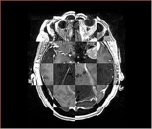
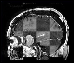
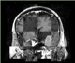
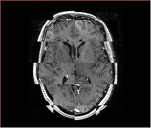
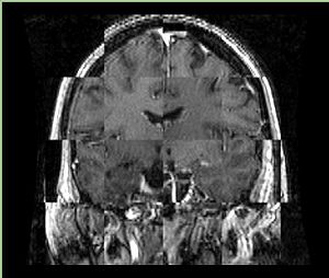
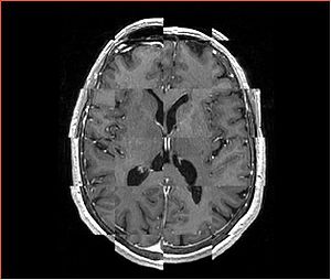
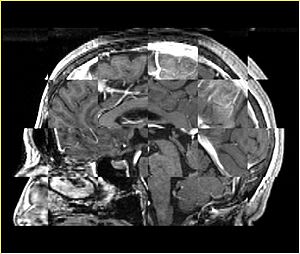
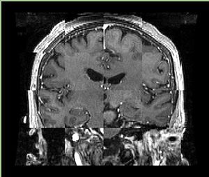
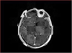
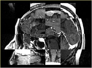
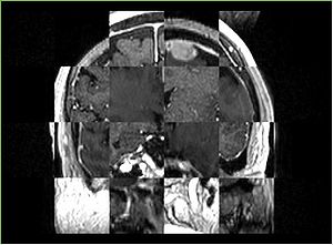
- Checkerboard appearance of unregistered images for the representative data of interest
- Case01
- Case03
- Case04
- Case10
Environment
All registration methods were executed on george.bwh.harvard.edu. Where number of threads was possible to specify, it was set to 4.
Measures of success
- qualitative assessment: visually pleasing results
- quantitative assessment: something better than "visually pleasing" (TBD)
- minimum execution time to meet the application requirements: interactive quantification of tumor growth
Methods
Registration
RegisterImages Slicer module
RigidRegistration Slicer module
BRAINSFit
- BRAINSFit on NITRC
- Technical details on InsightJournal
- Parameters:
${SLICER} --launch BRAINSFitExe \
  --fixedVolume ${TIME_POINT_1}  --movingVolume ${TIME_POINT_2} \
  --transformType Rigid --initializeTransformMode CenterOfHead \
  --numberOfIterations 200 --numberOfSamples 100000 --minimumStepSize 0.005 \
  --spatialScale 1000 --reproportionScale 1 \
  --outputTransform ${BF_TRANSFORM} --histogramMatch \
  --strippedOutputTransform ${BF_STRIPPED_TRANSFORM} \
  --patientID ANON --studyID ANON --outputVolumePixelType float \
  --backgroundFillValue 0 --maskProcessingMode ROIAUTO \
  --fixedVolumeTimeIndex 0 --movingVolumeTimeIndex 0 \
  --fixedVolumeOrigin 0,0,0 --movingVolumeOrigin 0,0,0 \
  --medianFilterSize 0,0,0 --permitParameterVariation 1,1,1 \
  --useCachingOfBSplineWeightsMode ON --useExplicitPDFDerivativesMode Whatever \
  --relaxationFactor 0.5 --maximumStepSize 0.2 --failureExitCode -1 \
  --fixedVolumeROI ${FIXED_IMAGE_ROI} --movingVolumeROI ${MOVING_IMAGE_ROI} \
  --numberOfHistogramBins 50 --numberOfMatchPoints 10 --debugNumberOfThreads 4
Note: all parameters are the default settings except numberOfIterations (default 1500), maskProcessingMode (default NOMASK).
FSL FLIRT
- FSL FLIRT
- Technical details
- parameters:
flirt -in ${TIME_POINT_2} -ref ${TIME_POINT_1} -omat ${FLIRT_TRANSFORM} -out ${RESAMPLED_IMAGE} -dof 6 -v
- execution time: ~4 min
