Rhesus Subcortical Atlas
From NAMIC Wiki
Home < Rhesus Subcortical Atlas
Objective:
- Develop an atlas of subcortical structures from normal subject images.
Progress:
- We are currently developing a subcortical atlas following the methods outlined in
M. Styner, R. Knickmeyer, S. Joshi, C. Coe, S. J. Short, and J. Gilmore. Automatic brain segmentation in rhesus monkeys. Proc SPIE Medical Imaging Conference, Proc SPIE Vol 6512 Medical Imaging 2007, pp 65122L-1 - 65122L-8.
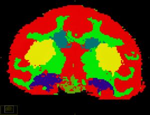
- Below is a snap shot of subcortical segmention of one of the subjects. The hippocampus, caudate and putamen are clearly visible (marked in blue, yellow and indigo) amidst the gray matter(marked in red) and WM (marked in green).
Key Investigators:
- Wake Forest: Bob Kraft, Jim Daunais
- Virginia Tech: Chris Wyatt, Vidya Rajagopalan
Links:
