Uncategorized files
From NAMIC Wiki
Showing below up to 50 results in range #1,551 to #1,600.
View (previous 50 | next 50) (20 | 50 | 100 | 250 | 500)
 Both1-cut.png 744 × 362; 66 KB
 BottomDeselectedIndicatorAndIconBlank.png 21 × 36; 354 bytes
 BottomDisabledIndicatorAndIconBlank.png 21 × 36; 373 bytes
 BottomSelectedCheckIndicatorAndIconBlank.png 21 × 36; 414 bytes
 BottomSelectedRadioIndicatorAndIconBlank.png 21 × 36; 390 bytes
 Bottom view patient3 time1.png 459 × 388; 115 KB
 Bottom view patient3 time2.png 459 × 385; 117 KB
 Box fmap.png 202 × 158; 22 KB
 Box fmap lp.png 197 × 149; 35 KB
 Box mag.jpg 199 × 151; 3 KB
 Box mag.png ; 15 KB
- Box mag1.png ; 15 KB
 Box susc.png 195 × 147; 4 KB
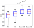 BoxplotDice.png 828 × 678; 51 KB
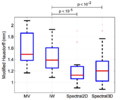 BoxplotHausdorff.png 812 × 676; 51 KB
 Brain-flat.PNG 390 × 400; 152 KB
 Brain-seg-utah.png 1,412 × 1,676; 20 KB
- Brain-seg-utah.tif 1,412 × 1,676; 109 KB
 Brain.PNG 370 × 415; 178 KB
 Brain.png 320 × 207; 10 KB
 BrainAfter.png 1,326 × 860; 248 KB
- BrainAtlasClassifier.zip ; 39.9 MB
- BrainAtlasClassifier2.zip ; 40.31 MB
 BrainBefore.png 1,120 × 857; 154 KB
 BrainColor-logo.png 303 × 141; 30 KB
 BrainDevelopment.jpg 177 × 140; 6 KB
 BrainH.png 200 × 175; 46 KB
 BrainLab-BioImageSuite-Slicer.jpg 1,280 × 1,024; 205 KB
 BrainMRI.compass2.png 165 × 154; 33 KB
 BrainShiftDiagram.png 1,565 × 471; 192 KB
 BrainTissueClassifiers BouixNeuroimage2007.pdf 0 × 0; 3.55 MB
 BrainVentriclesAndLesions-c.png 1,020 × 632; 150 KB
 BrainVentriclesAndLesions.png 855 × 776; 182 KB
- Brain Atlas Tutorial.ppt ; 7.01 MB
- Brain Atlas Tutorial07.ppt ; 3.92 MB
- Brain Atlas Tutorial08.ppt ; 3.19 MB
 Brain Mask Prediction Based on MRI Skin Data.png 1,920 × 1,027; 226 KB
 Brain sag2.jpg 1,300 × 904; 175 KB
 Brain sag 3d.jpg 698 × 502; 135 KB
 Brain sag axial.jpg 657 × 905; 130 KB
 Brain sag coronal.jpg 653 × 903; 133 KB
 Braindevelopment.jpg 700 × 490; 26 KB
 Braindevelopment qa.jpg 614 × 507; 39 KB
 Brainflat1.gif 303 × 244; 35 KB
 Brainflat2.gif 246 × 245; 31 KB
 Brainseg1.jpg 753 × 563; 52 KB
 Brainstem contours 3D.png 1,310 × 795; 928 KB
 Brainstem contours on different images.png 952 × 824; 887 KB
 Brainverse.png 1,856 × 1,306; 171 KB
 Branchsplitting.png 856 × 453; 79 KB


