File list
From NAMIC Wiki
This special page shows all uploaded files.
| Date | Name | Thumbnail | Size | User | Description | Versions |
|---|---|---|---|---|---|---|
| 12:34, 10 January 2014 | OverlaidView3.png (file) |  |
1.24 MB | Ayamada | 1 | |
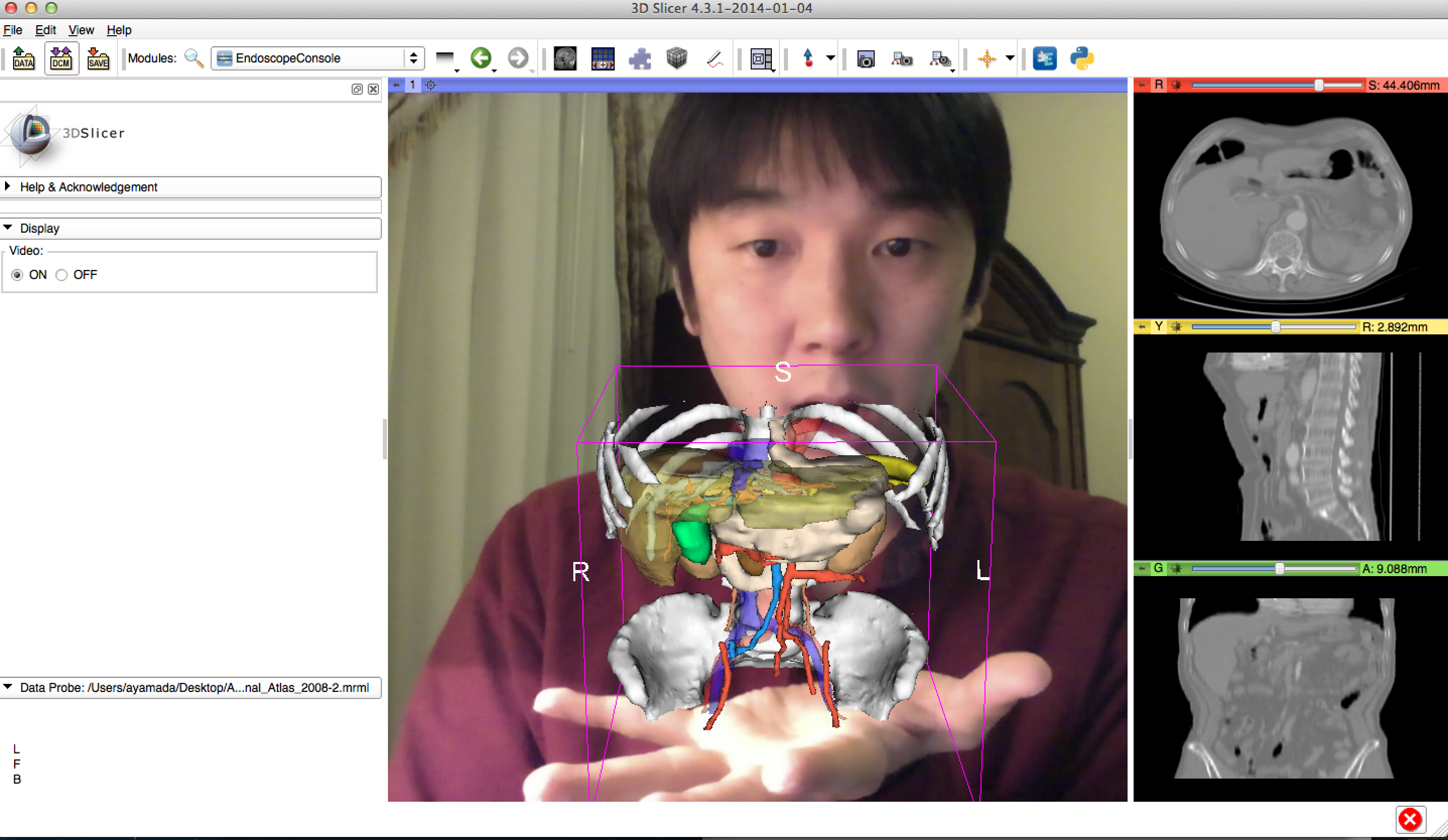
| 12:27, 10 January 2014 | OverlaidViewer2.png (file) |  |
1.75 MB | Ayamada | 1 | |
| 12:17, 10 January 2014 | OverlayedViewer.png (file) |  |
1.13 MB | Ayamada | 1 | |
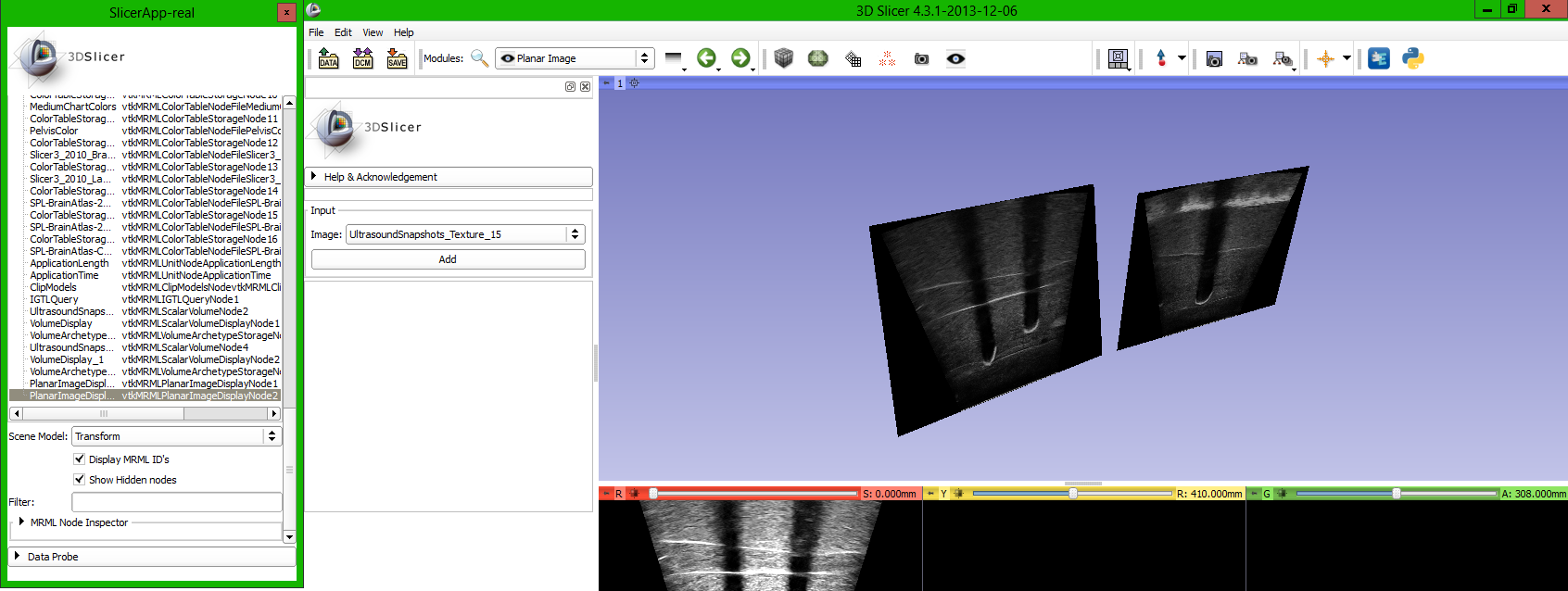
| 10:48, 10 January 2014 | PlanarImages 1.png (file) |  |
204 KB | Lasso | 1 | |
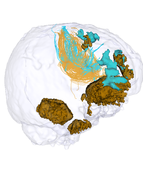
| 07:15, 10 January 2014 | Acute vs Chronic GenuBodyCC fibers and lesions 2.png (file) |  |
199 KB | Anuja | 1 | |
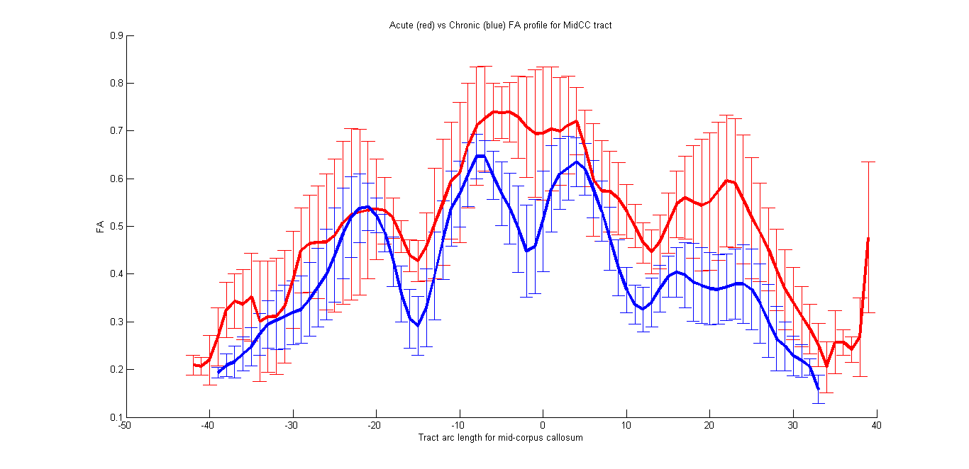
| 07:02, 10 January 2014 | Plot acute midcc.png (file) |  |
17 KB | Anuja | 1 | |
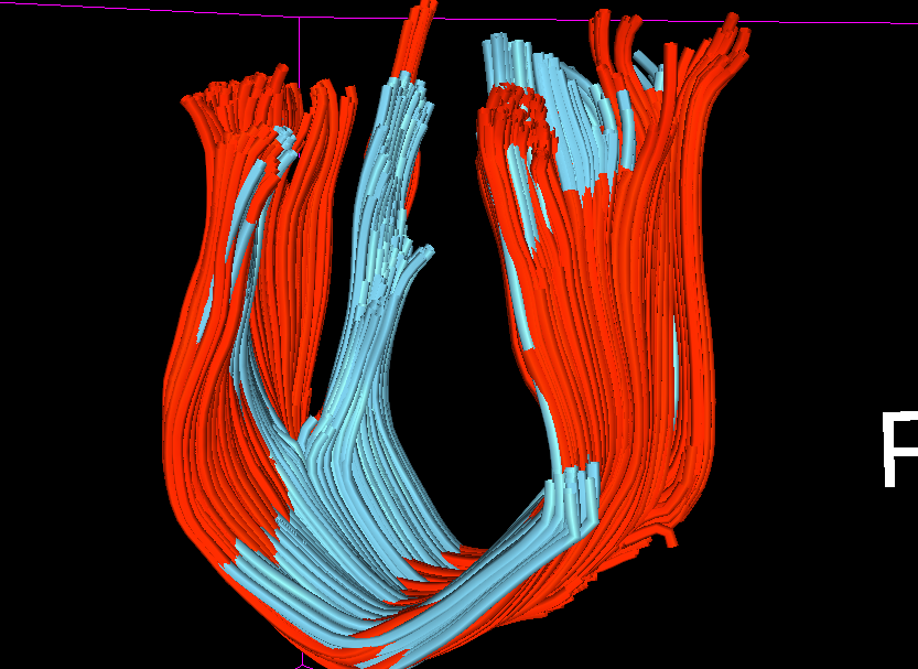
| 07:01, 10 January 2014 | Midcc2.png (file) |  |
281 KB | Anuja | 1 | |
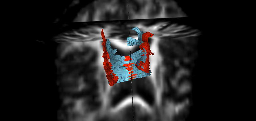
| 07:01, 10 January 2014 | Midcc.png (file) |  |
126 KB | Anuja | 1 | |
| 07:01, 10 January 2014 | Coreg chronic to acute2.png (file) |  |
62 KB | Anuja | 1 | |
| 07:00, 10 January 2014 | Coreg chronic to acute.png (file) |  |
91 KB | Anuja | 1 | |
| 06:52, 10 January 2014 | Cattura1.JPG (file) |  |
52 KB | PietroNardelli | Chest labelmap after user selection | 1 |
| 06:51, 10 January 2014 | Cattura.JPG (file) |  |
62 KB | PietroNardelli | Chest labelmap | 1 |
| 06:47, 10 January 2014 | Chronic redorig blueQC.png (file) |  |
135 KB | Anuja | 1 | |
| 06:47, 10 January 2014 | Acute redorig blueqc.png (file) |  |
201 KB | Anuja | 1 | |
| 06:37, 10 January 2014 | ChronicQC dti2.png (file) |  |
491 KB | Anuja | 1 | |
| 06:37, 10 January 2014 | Chronic dti2.png (file) |  |
465 KB | Anuja | 1 | |
| 06:37, 10 January 2014 | Acute QCed dti1.png (file) |  |
840 KB | Anuja | 1 | |
| 06:36, 10 January 2014 | Acute dti1.png (file) |  |
421 KB | Anuja | 1 | |
| 06:00, 10 January 2014 | Aging-1.jpg (file) |  |
184 KB | Andrei | 1 | |
| 05:51, 10 January 2014 | TBI-demyelination-2.jpg (file) |  |
143 KB | Andrei | 1 | |
| 05:51, 10 January 2014 | TBI-demyelination-1.jpg (file) |  |
192 KB | Andrei | 1 | |
| 05:47, 10 January 2014 | AcSeg final seg normal tissue classes.jpg (file) |  |
530 KB | Bowang | 1 | |
| 05:47, 10 January 2014 | AcSeg final alphamap.jpg (file) |  |
477 KB | Bowang | 1 | |
| 05:47, 10 January 2014 | AcSeg activelearning.jpg (file) |  |
450 KB | Bowang | 1 | |
| 05:47, 10 January 2014 | AcSeg selftraining.jpg (file) |  |
375 KB | Bowang | 1 | |
| 05:46, 10 January 2014 | AcSeg initial box.jpg (file) |  |
398 KB | Bowang | 1 | |
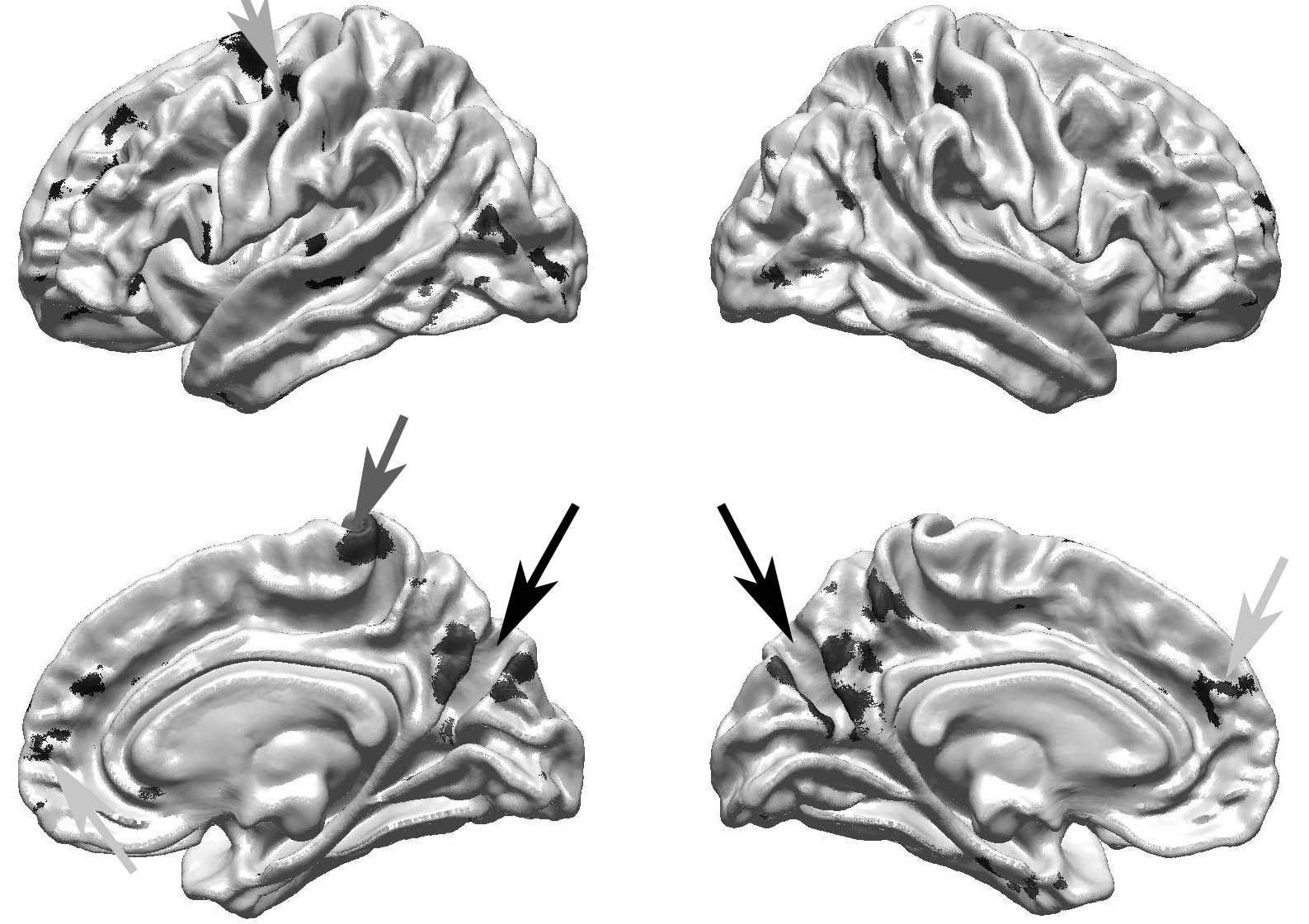
| 05:43, 10 January 2014 | TBI atrophy medial.jpg (file) |  |
29 KB | Andrei | 1 | |
| 05:42, 10 January 2014 | TBI atrophy lateral.jpg (file) |  |
29 KB | Andrei | 1 | |
| 05:42, 10 January 2014 | TBI atrophy inferior.jpg (file) |  |
25 KB | Andrei | 1 | |
| 05:39, 10 January 2014 | TBI atrophy inferior.tif (file) | 1.06 MB | Andrei | 1 | ||
| 05:38, 10 January 2014 | TBI atrophy medial.tif (file) | 1.06 MB | Andrei | 1 | ||
| 05:38, 10 January 2014 | TBI atrophy lateral.tif (file) | 1.06 MB | Andrei | 1 | ||
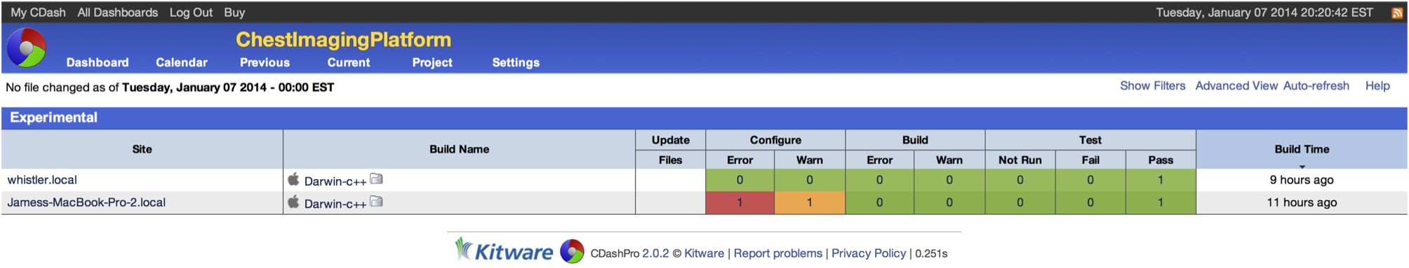
| 05:18, 10 January 2014 | CIP CDash.jpg (file) | 93 KB | Jcross186 | 1 | ||
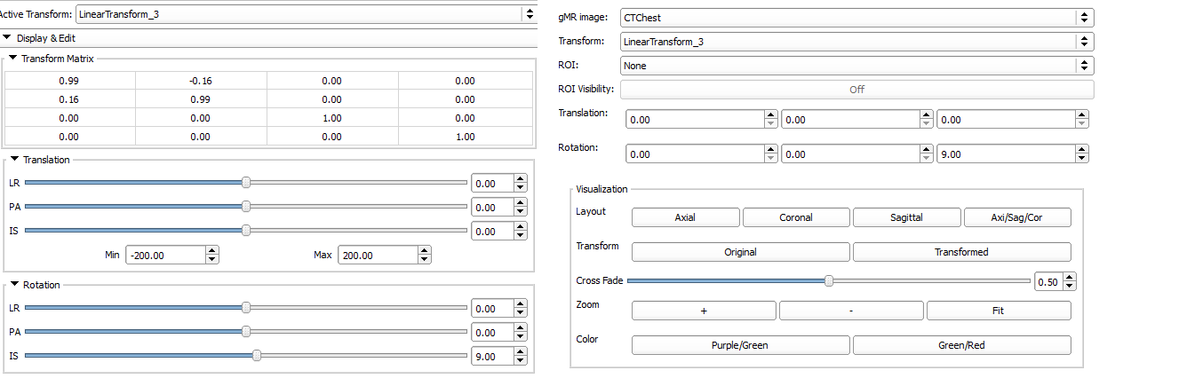
| 04:02, 10 January 2014 | Mrgrt interactive reg.png (file) |  |
23 KB | Wangk | 1 | |
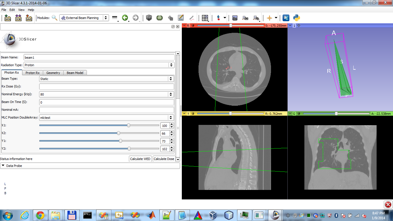
| 03:49, 10 January 2014 | SlicerRT MLC beam visualization.png (file) |  |
331 KB | Wangk | 1 | |
| 03:34, 10 January 2014 | 51839 forward vs backward.gif (file) |  |
4.5 MB | Jfishbaugh | 1 | |
| 03:25, 10 January 2014 | 51389 bw.gif (file) |  |
2.18 MB | Jfishbaugh | 1 | |
| 03:23, 10 January 2014 | 51389 fw.gif (file) |  |
2.22 MB | Jfishbaugh | 1 | |
| 03:22, 10 January 2014 | MRAFusion Step2.png (file) |  |
616 KB | Sbengali | 2 | |
| 03:20, 10 January 2014 | MRAFusion Step1.png (file) |  |
526 KB | Sbengali | 1 | |
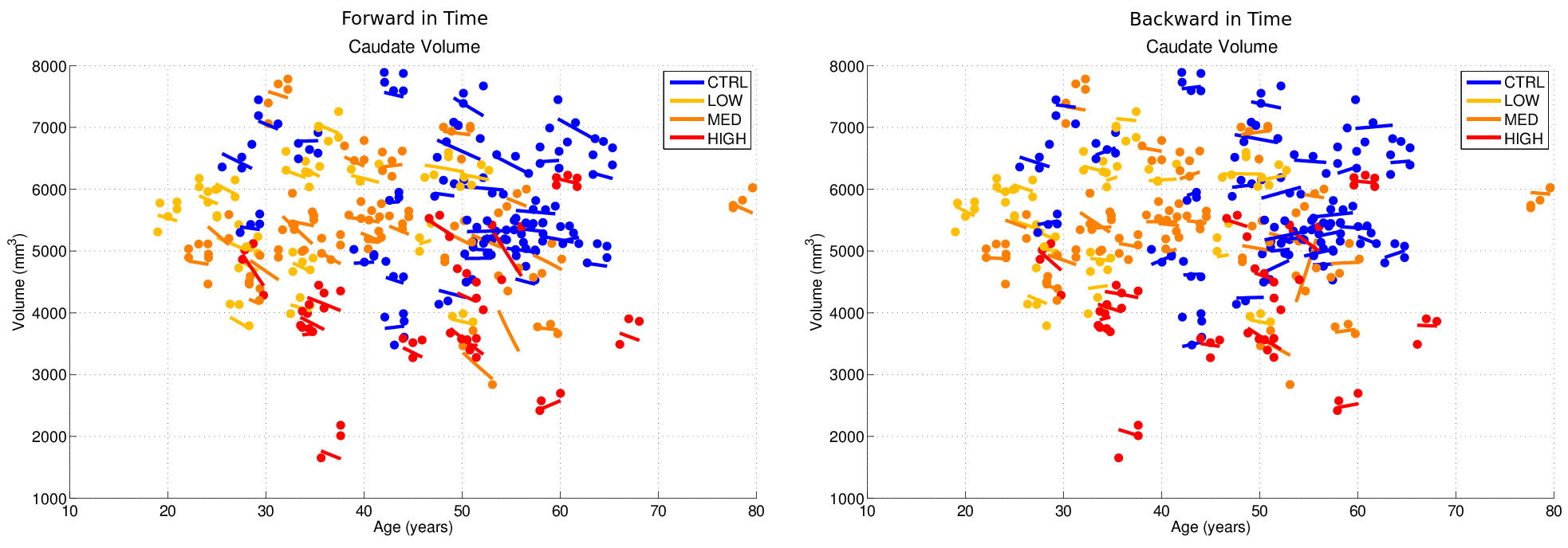
| 03:06, 10 January 2014 | Caudate vol compare fw bw.png (file) |  |
110 KB | Jfishbaugh | 1 | |
| 02:53, 10 January 2014 | First to last.png (file) |  |
220 KB | Jfishbaugh | 1 | |
| 02:53, 10 January 2014 | Registered shape.png (file) |  |
345 KB | Jfishbaugh | 1 | |
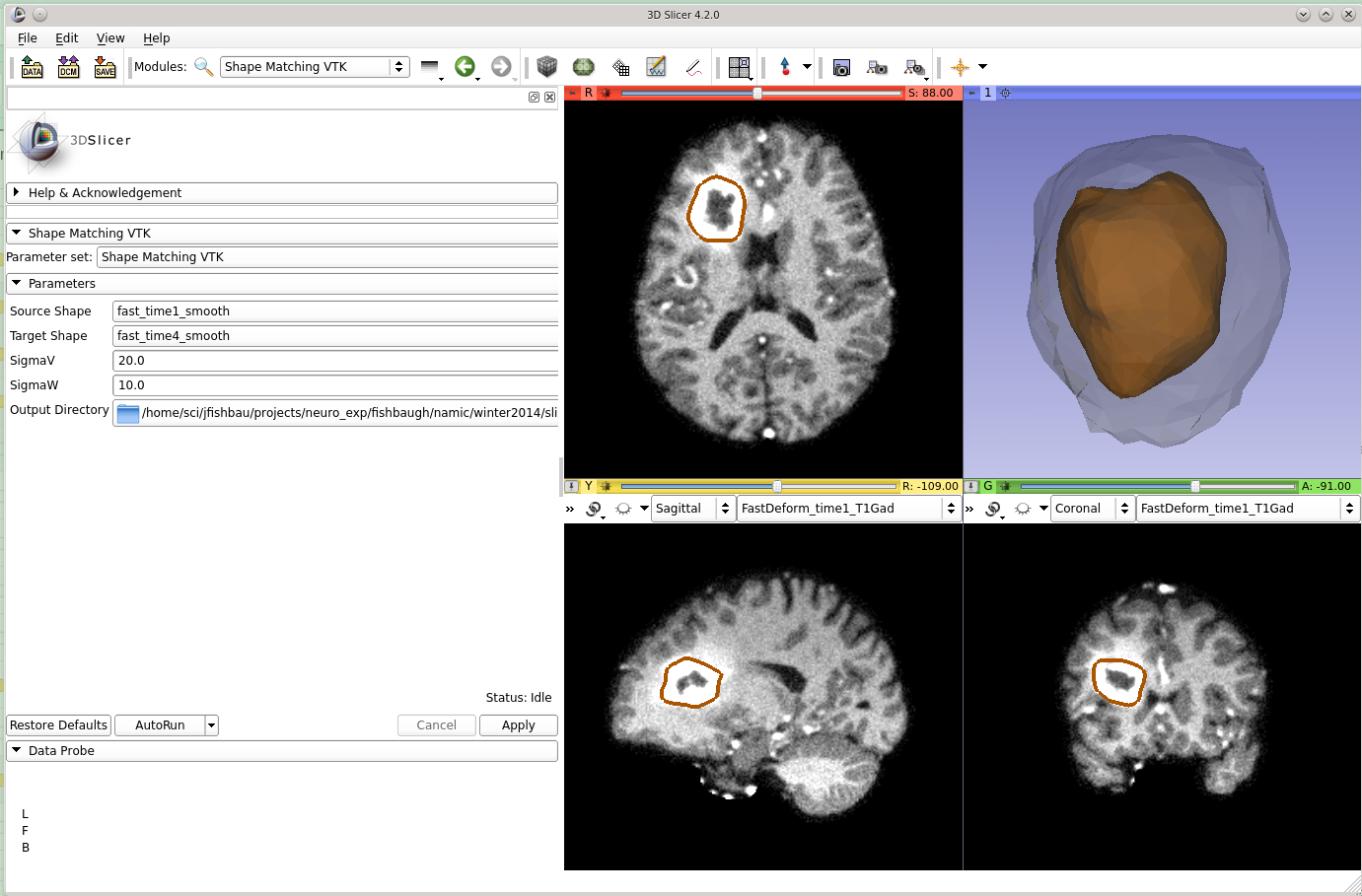
| 02:51, 10 January 2014 | Shape matching interface.png (file) |  |
359 KB | Jfishbaugh | 1 | |
| 02:48, 10 January 2014 | Init data.png (file) |  |
360 KB | Jfishbaugh | 1 | |
| 02:15, 10 January 2014 | LAFibrosisVis2.png (file) |  |
74 KB | Sbengali | 1 | |
| 02:07, 10 January 2014 | LAFIbrosisVis.png (file) |  |
74 KB | Sbengali | 1 | |
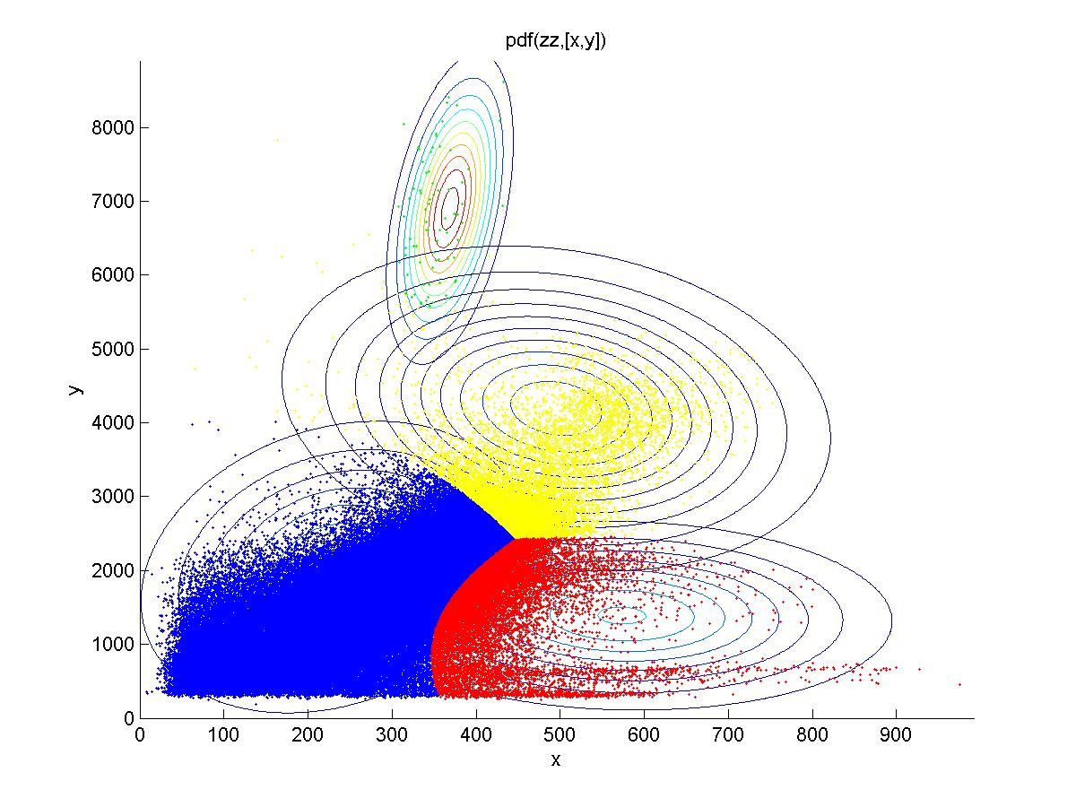
| 00:55, 10 January 2014 | Stroke lesion mixture model.jpg (file) |  |
160 KB | Rameshvs | Different tissue classes found using Gaussian mixture models for FLAIR (x) and DWI (y). Yellow corresponds to (oversegmented) stroke lesion, blue corresponds to normal tissue, and red and green are artifacts and white matter hyperintensity respectively. | 1 |
| 00:35, 10 January 2014 | NAMIC AHM 2014 van horn.pdf (file) | 7.25 MB | Jvanhorn | 1 | ||
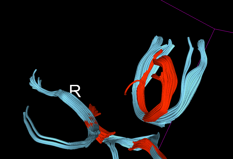
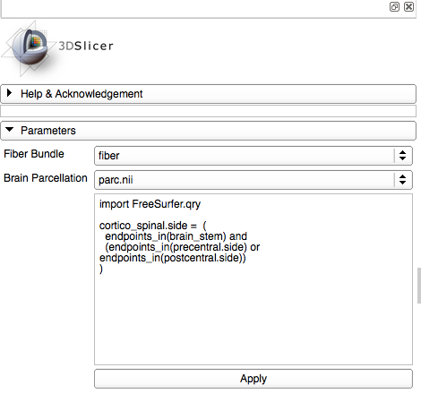
| 00:25, 10 January 2014 | WMQLModuleSampleOutput1.png (file) |  |
33 KB | Petersv | 1 |