Difference between revisions of "2009 Summer Project Week Meningioma growth simulation"
| Line 46: | Line 46: | ||
</div> | </div> | ||
</div> | </div> | ||
| + | |||
| + | <h3>Meningioma simulation parameter setup</h3> | ||
| + | |||
| + | The following xml configuration was used for initial evaluation: | ||
| + | <pre> | ||
| + | <?xml version="1.0"?> | ||
| + | <tumor-simulation-parameters> | ||
| + | <dataset-name>SimMeningioma001</dataset-name> | ||
| + | <input-directory>/scratch/prastawa/TumorSim/TumorSimInput</input-directory> | ||
| + | <output-directory>/scratch/prastawa/TumorSim/Meningioma001</output-directory> | ||
| + | <deformation-seed>/scratch/prastawa/TumorSim/seed_m1.mha</deformation-seed> | ||
| + | <deformation-iterations>8</deformation-iterations> | ||
| + | <infiltration-iterations>4</infiltration-iterations> | ||
| + | <infiltration-body-force-iterations>4</infiltration-body-force-iterations> | ||
| + | <deformation-initial-pressure>5</deformation-initial-pressure> | ||
| + | <deformation-kappa>100</deformation-kappa> | ||
| + | <infiltration-time-step>0.5</infiltration-time-step> | ||
| + | <infiltration-body-force-coefficient>8</infiltration-body-force-coefficient> | ||
| + | <contrast-enhancement-type>uniform</contrast-enhancement-type> | ||
| + | </tumor-simulation-parameters> | ||
| + | </pre> | ||
<h3>References</h3> | <h3>References</h3> | ||
Revision as of 14:59, 10 June 2009
Home < 2009 Summer Project Week Meningioma growth simulation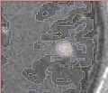
Tumor sumulation iteration 1

... iteration 2
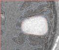
... iteration 3


Key Investigators
- BWH: Andriy Fedorov, Ron Kikinis
- Utah: Marcel Prastawa
Objective
Meningiomas are primary brain tumors that grow from the cells of meninges. Such tumors are typically benign, and are not invasive. In this project we will investigate the use of the tumor growth simulation tool, TumorSim, for the purposes of modeling the growth of meningioma. Our primary objective is to tune the parameters of the simulation to have maximally realistic appearance of contrast-enhanced tumor mass, realistic deformation of the surrounding tissue, and to evaluate the capabilities of the tumor simulator in modeling meningiomas of various morphology.
Approach, plan
TumorSim software allows to specify the following simulation parameters:
- deformation parameters
- number of iterations
- initial pressure
- kappa
- infiltration parameters
- number of iterations
- number of body force iterations
- time step
- body force coefficient
- contrast enhancement type: none, ring or uniform
We will analyze contrast-enhanced MRIs of meningiomas of different sizes, located in different areas of brain, and will attempt to develop a set of recommendations for initializations of the parameters to mimic these tumors.
Additional practical considerations:
- how can the level of noise be controlled?
- what is the influence of the initialization label size on the tumor shape?
- tumor-brain interface appearance and correspondence with the tumor label
Progress
Meningioma simulation parameter setup
The following xml configuration was used for initial evaluation:
<?xml version="1.0"?>
<tumor-simulation-parameters>
<dataset-name>SimMeningioma001</dataset-name>
<input-directory>/scratch/prastawa/TumorSim/TumorSimInput</input-directory>
<output-directory>/scratch/prastawa/TumorSim/Meningioma001</output-directory>
<deformation-seed>/scratch/prastawa/TumorSim/seed_m1.mha</deformation-seed>
<deformation-iterations>8</deformation-iterations>
<infiltration-iterations>4</infiltration-iterations>
<infiltration-body-force-iterations>4</infiltration-body-force-iterations>
<deformation-initial-pressure>5</deformation-initial-pressure>
<deformation-kappa>100</deformation-kappa>
<infiltration-time-step>0.5</infiltration-time-step>
<infiltration-body-force-coefficient>8</infiltration-body-force-coefficient>
<contrast-enhancement-type>uniform</contrast-enhancement-type>
</tumor-simulation-parameters>
References
- M.Prastawa, E.Bullitt, and G.Gerig. Simulation of Brain Tumors in MR Images for Evaluation of Segmentation Efficacy. Medical Image Analysis 13(2):297-311, 2009 [1]
- TumorSim: Simulated Brain Tumor MRI Software and data
- Wikipedia: Meningioma