Difference between revisions of "2009 Summer Project Week Project Segmentation of Muscoskeletal Images"
m (Fix MediaWiki formatting issue discovered while converting to GitHub Flavored Markdown using pandoc (via https://github.com/outofcontrol/mediawiki-to-gfm)) Tag: 2017 source edit |
|||
| (17 intermediate revisions by 4 users not shown) | |||
| Line 1: | Line 1: | ||
__NOTOC__ | __NOTOC__ | ||
| − | <gallery> | + | <gallery widths="400px" perrow="6"> |
| − | Image:PW2009-v3.png|[[2009_Summer_Project_Week| | + | Image:PW2009-v3.png|[[2009_Summer_Project_Week#Projects|Projects List]] |
| − | Image: | + | Image:Knee.jpg| Knee MRI Image |
| − | Image: | + | Image:LabelMap Femur.jpg|Filled Femur Label Map |
| + | Image:Femur Patella Tibia.jpg|EM Segmented Output | ||
| + | Image:Logo_simbios.gif | ||
| + | Image:64 58 Affine 350 Iterations.jpeg | 64 Affine Registered on 58 | ||
| + | Image:64 58 PipeLinedAffine Registration 200 Iterations.JPG | 64 Pipeline Affine Registered on 58 | ||
| + | Image:64 58 BSpline 210 Iterations.JPG | 64 Bspline Registered on 58 | ||
| + | Image:42 Masked.jpg | Masked 42 MRI | ||
| + | Image:64 Masked.jpg | Masked 64 MRI | ||
| + | Image:42 BSpline To 64 DifferenceAfter.jpg | Registered Image Difference of 42 and 64 | ||
| + | Image:42 Registered To 64.jpg | BSpline Registered Output of 42 and 64 | ||
</gallery> | </gallery> | ||
==Key Investigators== | ==Key Investigators== | ||
| − | * | + | * Stanford: Harish Doddi, Saikat Pal, Scott Delp |
| − | * | + | * Harvard: Ron Kikinis |
| + | * Steve Pieper, Isomics, Inc. | ||
| + | * Luis Ibanez, Kitware, Inc. | ||
| − | |||
<div style="width: 27%; float: left; padding-right: 3%;"> | <div style="width: 27%; float: left; padding-right: 3%;"> | ||
<h3>Objective</h3> | <h3>Objective</h3> | ||
| − | + | The aim of this project is to develop an automatic/semi-automatic methodology to convert whole body imaging datasets into three-dimensional models for neuromuscular biomechanics and finite element simulations. | |
| + | |||
| + | Specific Aims | ||
| + | * Explore the segmentation techniques for knee MRI datasets with special focus on patella, femur and tibia bones. | ||
</div> | </div> | ||
| − | <div style="width: | + | <div style="width: 30%; float: left; padding-right: 3%;"> |
<h3>Approach, Plan</h3> | <h3>Approach, Plan</h3> | ||
| − | Our | + | We are working on understanding the capabilities of RegisterImage module in Slicer to apply image-to-image registration on knee datasets. We are implementing masking algorithms to isolate specific knee bones, and perform parameter exploration to evaluate the sensitivity of registered images to input parameters. We are exploring the feasibility of applying python ICP-based registration algorithms to directly morph a surface model to a target image geometry. |
| + | |||
| + | Our goals for the project week are: | ||
| + | * Implement an algorithm to acquire masked regions of interest from MR datasets. | ||
| + | * Perform and evaluate results from a parameter space exploration study of RegisterImages Batchmake module on knee dataset. | ||
| + | * Resolve issues in building Python modules from slicer source code. | ||
| + | * Explore possibility of model-to-image registration using existing atlas (.vtk, .stl) to a target image using Python ICP Registration module. | ||
| − | |||
</div> | </div> | ||
| − | <div style="width: | + | <div style="width: 27%; float: left;"> |
<h3>Progress</h3> | <h3>Progress</h3> | ||
| − | + | * Investigated the masking algorithm and completed the module to mask and register images. Need to explore the parameters technique for this code. | |
| + | * Resolved issues in building Python | ||
| + | * Cluster set up completed | ||
| + | * Converted Model to masked label map volume to further a masked label volume. Now we have the problem of registering label map volume to an MRI image. | ||
| + | |||
</div> | </div> | ||
| − | |||
| − | |||
| − | |||
| − | |||
| − | |||
| − | |||
| − | |||
| − | |||
Latest revision as of 19:54, 11 April 2023
Home < 2009 Summer Project Week Project Segmentation of Muscoskeletal Images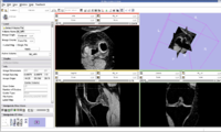
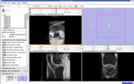
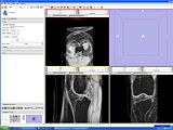
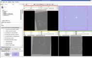
Knee MRI Image
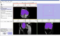
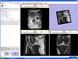
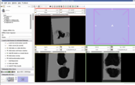
Filled Femur Label Map
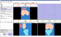
EM Segmented Output

64 Affine Registered on 58
64 Pipeline Affine Registered on 58
64 Bspline Registered on 58
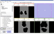
Masked 42 MRI
Masked 64 MRI
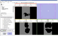
Registered Image Difference of 42 and 64
BSpline Registered Output of 42 and 64


Key Investigators
- Stanford: Harish Doddi, Saikat Pal, Scott Delp
- Harvard: Ron Kikinis
- Steve Pieper, Isomics, Inc.
- Luis Ibanez, Kitware, Inc.
Objective
The aim of this project is to develop an automatic/semi-automatic methodology to convert whole body imaging datasets into three-dimensional models for neuromuscular biomechanics and finite element simulations.
Specific Aims
- Explore the segmentation techniques for knee MRI datasets with special focus on patella, femur and tibia bones.
Approach, Plan
We are working on understanding the capabilities of RegisterImage module in Slicer to apply image-to-image registration on knee datasets. We are implementing masking algorithms to isolate specific knee bones, and perform parameter exploration to evaluate the sensitivity of registered images to input parameters. We are exploring the feasibility of applying python ICP-based registration algorithms to directly morph a surface model to a target image geometry.
Our goals for the project week are:
- Implement an algorithm to acquire masked regions of interest from MR datasets.
- Perform and evaluate results from a parameter space exploration study of RegisterImages Batchmake module on knee dataset.
- Resolve issues in building Python modules from slicer source code.
- Explore possibility of model-to-image registration using existing atlas (.vtk, .stl) to a target image using Python ICP Registration module.
Progress
- Investigated the masking algorithm and completed the module to mask and register images. Need to explore the parameters technique for this code.
- Resolved issues in building Python
- Cluster set up completed
- Converted Model to masked label map volume to further a masked label volume. Now we have the problem of registering label map volume to an MRI image.