Difference between revisions of "2009 Summer Project Week WML SEgmentation"
Medcomparts (talk | contribs) |
|||
| (4 intermediate revisions by 2 users not shown) | |||
| Line 2: | Line 2: | ||
<gallery> | <gallery> | ||
Image:PW2009-v3.png|[[2009_Summer_Project_Week#Projects|Projects List]] | Image:PW2009-v3.png|[[2009_Summer_Project_Week#Projects|Projects List]] | ||
| + | Image:itk_wmls_pipeline.png| Pipeline of WML segmentation | ||
Image:UNCWMLSegmentation.png|One training dataset (T1, T2, PD, FLAIR images and wml segmentation) | Image:UNCWMLSegmentation.png|One training dataset (T1, T2, PD, FLAIR images and wml segmentation) | ||
Image:itk_wmls.png| One testing dataset and segmentation result | Image:itk_wmls.png| One testing dataset and segmentation result | ||
| Line 15: | Line 16: | ||
<h3>Objective</h3> | <h3>Objective</h3> | ||
| − | We will continue | + | We will continue developing and testing the white matter lesion segmentation algorithm implemented using ITK. The goal is to have an initial version ready by the end of the week that can be distributed within NA-MIC community for more extensive testing. |
</div> | </div> | ||
| Line 29: | Line 30: | ||
<h3>Progress</h3> | <h3>Progress</h3> | ||
| − | Since winter project week in Utah, we have developed/implemented WML segmentation algorithm using ITK classes. Subtasks implemented | + | Since winter project week in Utah, we have developed/implemented a WML segmentation algorithm using ITK classes. Subtasks implemented include: (1) a skull stripping algorithm working on T1 weighted images; (2) a fuzzy clustering algorithm for tissue segmentation; (3) a parametric model for gain field correction. All of these subtasks are implemented by using ITK. The training step uses AdaBoost and the segmenation step uses a support vector machine. |
</div> | </div> | ||
| Line 35: | Line 36: | ||
==References== | ==References== | ||
| − | * Zhiqiang Lao, Dinggang Shen, Dengfeng Liu, Abbas F Jawad, Elias R Melhem, Lenore J Launer, Nick R Bryan, Christos Davatzikos, | + | * Zhiqiang Lao, Dinggang Shen, Dengfeng Liu, Abbas F Jawad, Elias R Melhem, Lenore J Launer, Nick R Bryan, Christos Davatzikos, Computer-Assisted Segmentation of White Matter Lesions in 3D MR images, Using Pattern Recognition, Academic Radiology, 15(3):300-313, March 2008. |
| + | [http://www.academicradiology.org/article/S1076-6332(07)00583-1/abstract] | ||
Latest revision as of 14:45, 13 August 2009
Home < 2009 Summer Project Week WML SEgmentation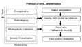
Pipeline of WML segmentation

One training dataset (T1, T2, PD, FLAIR images and wml segmentation)
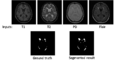
One testing dataset and segmentation result


Key Investigators
- UNC: Minjeong Kim, Dinggang Shen
- GE : Xiaodong Tao, Jim Miller
Objective
We will continue developing and testing the white matter lesion segmentation algorithm implemented using ITK. The goal is to have an initial version ready by the end of the week that can be distributed within NA-MIC community for more extensive testing.
Approach, Plan
We will develop a Slicer module for the white matter lesion segmentation algorithm. Base line results and test will be generated.
Progress
Since winter project week in Utah, we have developed/implemented a WML segmentation algorithm using ITK classes. Subtasks implemented include: (1) a skull stripping algorithm working on T1 weighted images; (2) a fuzzy clustering algorithm for tissue segmentation; (3) a parametric model for gain field correction. All of these subtasks are implemented by using ITK. The training step uses AdaBoost and the segmenation step uses a support vector machine.
References
- Zhiqiang Lao, Dinggang Shen, Dengfeng Liu, Abbas F Jawad, Elias R Melhem, Lenore J Launer, Nick R Bryan, Christos Davatzikos, Computer-Assisted Segmentation of White Matter Lesions in 3D MR images, Using Pattern Recognition, Academic Radiology, 15(3):300-313, March 2008.