Difference between revisions of "2011 Summer Project Week NerveSeg"
| (11 intermediate revisions by the same user not shown) | |||
| Line 1: | Line 1: | ||
| − | Nerve | + | <h1>Segmentation of Nerve Bundles and Ganglia in Spine MRI</h1> |
| − | |||
__NOTOC__ | __NOTOC__ | ||
| Line 15: | Line 14: | ||
Image:NerveSeg-res-scr2.png|Figure 4 - Example_2: Complete render of nerve segmentations. | Image:NerveSeg-res-scr2.png|Figure 4 - Example_2: Complete render of nerve segmentations. | ||
Image:NerveSeg-res-scr3.png|Figure 5 - Example_2: Another View. | Image:NerveSeg-res-scr3.png|Figure 5 - Example_2: Another View. | ||
| − | + | Image:NerveSeg-mapo.png|Figure 6 - Example_3: Map of automatically segmented nerves. Circled in red are areas of sudden changes. | |
| − | |||
| − | |||
| − | |||
| − | |||
| − | Image:NerveSeg- | ||
| − | |||
</gallery> | </gallery> | ||
| Line 38: | Line 31: | ||
<div style="width: 27%; float: left; padding-right: 3%;"> | <div style="width: 27%; float: left; padding-right: 3%;"> | ||
| − | |||
<h3>Approach, Plan</h3> | <h3>Approach, Plan</h3> | ||
| − | Currently we use a particle-filter tracking approach for segmenting the nerves. The algorithm is given a seed point, preferably somewhere in the spine. The particles are tubes following Bézier curves (and hence forming a B-spline track). The dynamics model encourages continuity and smoothness. The image likelihood model compares gradient fields and intensities of predicted patches with image observations to evaluate a posterior distribution of the particles' importance. While we can currently usually track the nerve cores, usually fully throughout the vertebral canal, the algorithm does not delineate the full extent of the nerves, and behaves poorly on peripheral nerves outside of the vertebral canal. We will modify the likelihood function and parameters and concentrate on achieving this during the project week. | + | Currently we use a particle-filter tracking approach for segmenting the nerves. The algorithm is given a seed point, preferably somewhere in the spine. The particles are tubes following Bézier curves (and hence forming a B-spline track). The dynamics model encourages continuity and smoothness. The image likelihood model compares gradient fields and intensities of predicted patches with image observations to evaluate a posterior distribution of the particles' importance. While we can currently usually track the nerve cores, usually fully throughout the vertebral canal, the algorithm does not delineate the full extent of the nerves and has sudden changes, and behaves poorly on peripheral nerves outside of the vertebral canal. We will modify the likelihood function and parameters and concentrate on achieving this during the project week. |
</div> | </div> | ||
| Line 48: | Line 40: | ||
<h3>Progress</h3> | <h3>Progress</h3> | ||
| + | * Sudden changes in the segmentation can be ameliorated. In general, can now constrain the radius function to be smoother in the general (visual) sense not just in the technical mathematical sense, to avoiding sudden changes in the segmentation (which are simply areas with fast but smooth radius function change). | ||
| + | * With careful characterization of the newer radius function, we can segment some of the thinner peripheral nerves. This is still specific to the images explored, but it can be done. | ||
| + | * Full width estimation is hard as part of the current system, may have to be a separate extension. | ||
| + | |||
| + | Results are attached: | ||
| + | |||
| + | <gallery> | ||
| + | Image:NerveSeg-peripheral.png|Figure 7 - Example_4: Another example, following several nerve bundles. | ||
| + | Image:NerveSeg-mapp.png|Figure 8 - Example_4: A map of several automatically segmented nerves with herniated disc manually segmented. | ||
| + | </gallery> | ||
| Line 55: | Line 57: | ||
<div style="width: 97%; float: left;"> | <div style="width: 97%; float: left;"> | ||
| − | + | ==References== | |
| − | + | A.V. Dalca; Giovanna Danagoulian; Ron kikinis; Ehud Schmidt; Polina Golland. (2011) Segmentation of Nerve Bundles and Ganglia in Spine MRI using Particle Filters. To Appear in MICCAI 2011 | |
| − | |||
| − | |||
</div> | </div> | ||
Latest revision as of 14:01, 24 June 2011
Home < 2011 Summer Project Week NerveSegSegmentation of Nerve Bundles and Ganglia in Spine MRI
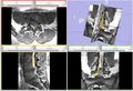
Figure 1 - Example of Manual Nerve Segmentation


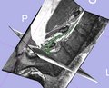
Figure 2 - Example_1: non-cleaned results.
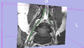
Figure 3 - Example_2: Automatic segmentation for multiple nerves. Right nerve is uncleaned, Left nerve is run with a clean-up post-processing step.
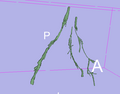
Figure 4 - Example_2: Complete render of nerve segmentations.
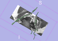
Figure 5 - Example_2: Another View.
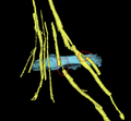
Figure 6 - Example_3: Map of automatically segmented nerves. Circled in red are areas of sudden changes.


Key Investigators
- MIT: Adrian Dalca, Polina Golland
- BWH: Giovanna Danagoulian, Ehud Schmidt
- SPL: Ron Kikinis
Objective
We are developing a nerve segmentation algorithm for the automatic isolation of nerves and nerve ganglia inside the spinal sack and out through the vertebrae in new MR Myelography images. Current progress can track the core of a Nerve.
Approach, Plan
Currently we use a particle-filter tracking approach for segmenting the nerves. The algorithm is given a seed point, preferably somewhere in the spine. The particles are tubes following Bézier curves (and hence forming a B-spline track). The dynamics model encourages continuity and smoothness. The image likelihood model compares gradient fields and intensities of predicted patches with image observations to evaluate a posterior distribution of the particles' importance. While we can currently usually track the nerve cores, usually fully throughout the vertebral canal, the algorithm does not delineate the full extent of the nerves and has sudden changes, and behaves poorly on peripheral nerves outside of the vertebral canal. We will modify the likelihood function and parameters and concentrate on achieving this during the project week.
Progress
- Sudden changes in the segmentation can be ameliorated. In general, can now constrain the radius function to be smoother in the general (visual) sense not just in the technical mathematical sense, to avoiding sudden changes in the segmentation (which are simply areas with fast but smooth radius function change).
- With careful characterization of the newer radius function, we can segment some of the thinner peripheral nerves. This is still specific to the images explored, but it can be done.
- Full width estimation is hard as part of the current system, may have to be a separate extension.
Results are attached:

Figure 7 - Example_4: Another example, following several nerve bundles.
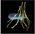
Figure 8 - Example_4: A map of several automatically segmented nerves with herniated disc manually segmented.


References
A.V. Dalca; Giovanna Danagoulian; Ron kikinis; Ehud Schmidt; Polina Golland. (2011) Segmentation of Nerve Bundles and Ganglia in Spine MRI using Particle Filters. To Appear in MICCAI 2011