Difference between revisions of "2011 Winter Project Week:Atrial Fibrillation"
| Line 14: | Line 14: | ||
==Key Investigators== | ==Key Investigators== | ||
| − | * | + | * Georgia Tech: Behnood Gholami, Yi Gao, and Allen Tannenbaum |
| − | |||
<div style="margin: 20px;"> | <div style="margin: 20px;"> | ||
| Line 21: | Line 20: | ||
<h3>Objective</h3> | <h3>Objective</h3> | ||
| − | |||
| − | |||
| − | |||
| − | |||
| + | Atrial fibrillation, a cardiac arrhythmia characterized by unsynchronized electrical activity in the atrial chambers of the heart, is a rapidly growing problem in modern societies. Electrical cardioversion and antiarrhythmic drugs are used to manage this condition, but suffer from low success rates and involve major side effects. In an alternative treatment, known as catheter ablation, specific parts of the left atrium are targeted for radio frequency ablation using an intracardiac catheter. Application of radio frequency energy to the cardiac tissue causes thermal injury (lesions), which in turn results into scar tissue. Successful ablation can eliminate, or isolate, the problematic sources of electrical activity and effectively cure atrial fibrillation. | ||
| + | Magnetic resonance imaging (MRI) has been used for both pre- and and post-ablation assessment of the atrial wall. MRI can aid in selecting the right candidate for the ablation procedure and assessing post-ablation scar formations. Image processing techniques can be used for automatic segmentation of the atrial wall, which facilitates an accurate statistical assessment of the region. As a first step towards the general solution to the computer-assisted segmentation of the left atrial wall, in this research we propose a shape-based image segmentation framework to segment the endocardial wall of the left atrium. | ||
| + | We are developing methods to segment the left atrial wall in delayed-enhanced MR imagery. | ||
</div> | </div> | ||
| Line 34: | Line 32: | ||
<h3>Approach, Plan</h3> | <h3>Approach, Plan</h3> | ||
| − | + | We plan to finalize a fully-automatic segmentation approach to identify the blood pool in MRAs. The approach uses the robust statistics segmentation framework developed earlier at Georgia Tech. | |
| − | |||
| − | |||
</div> | </div> | ||
| Line 43: | Line 39: | ||
<h3>Progress</h3> | <h3>Progress</h3> | ||
| − | |||
| Line 51: | Line 46: | ||
<div style="width: 97%; float: left;"> | <div style="width: 97%; float: left;"> | ||
| − | |||
| − | |||
| − | |||
| − | |||
| − | |||
| − | |||
| − | |||
| − | |||
| − | |||
| − | |||
==References== | ==References== | ||
| − | * | + | * Y. Gao, B. Gholami, R. MacLeod, J. Blauer, W. M. Haddad, and A. R. Tannenbaum, "Segmentation of the Endocardial Wall of the Left Atrium using Localized Region-Based Active Contours and Statistical Shape Learning," Proc. SPIE Med. Imag., San Diego, CA, vol. 7623, 76234Z-1, 2010. |
| − | |||
| − | |||
| − | |||
</div> | </div> | ||
Revision as of 19:21, 13 December 2010
Home < 2011 Winter Project Week:Atrial Fibrillation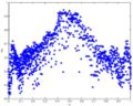
Scatter plot of the original FA data through the genu of the corpus callosum of a normal brain.
Regression of FA data; solid line represents the mean and dotted lines the standard deviation.


Instructions for Use of this Template
- Please create a new wiki page with an appropriate title for your project using the convention Project/<Project Name>
- Copy the entire text of this page into the page created above
- Link the created page into the list of projects for the project event
- Delete this section from the created page
- Send an email to tkapur at bwh.harvard.edu if you are stuck
Key Investigators
- Georgia Tech: Behnood Gholami, Yi Gao, and Allen Tannenbaum
Objective
Atrial fibrillation, a cardiac arrhythmia characterized by unsynchronized electrical activity in the atrial chambers of the heart, is a rapidly growing problem in modern societies. Electrical cardioversion and antiarrhythmic drugs are used to manage this condition, but suffer from low success rates and involve major side effects. In an alternative treatment, known as catheter ablation, specific parts of the left atrium are targeted for radio frequency ablation using an intracardiac catheter. Application of radio frequency energy to the cardiac tissue causes thermal injury (lesions), which in turn results into scar tissue. Successful ablation can eliminate, or isolate, the problematic sources of electrical activity and effectively cure atrial fibrillation.
Magnetic resonance imaging (MRI) has been used for both pre- and and post-ablation assessment of the atrial wall. MRI can aid in selecting the right candidate for the ablation procedure and assessing post-ablation scar formations. Image processing techniques can be used for automatic segmentation of the atrial wall, which facilitates an accurate statistical assessment of the region. As a first step towards the general solution to the computer-assisted segmentation of the left atrial wall, in this research we propose a shape-based image segmentation framework to segment the endocardial wall of the left atrium.
We are developing methods to segment the left atrial wall in delayed-enhanced MR imagery.
Approach, Plan
We plan to finalize a fully-automatic segmentation approach to identify the blood pool in MRAs. The approach uses the robust statistics segmentation framework developed earlier at Georgia Tech.
Progress
References
- Y. Gao, B. Gholami, R. MacLeod, J. Blauer, W. M. Haddad, and A. R. Tannenbaum, "Segmentation of the Endocardial Wall of the Left Atrium using Localized Region-Based Active Contours and Statistical Shape Learning," Proc. SPIE Med. Imag., San Diego, CA, vol. 7623, 76234Z-1, 2010.