Difference between revisions of "2011 Winter Project Week:SegEye"
From NAMIC Wiki
Ivan.kolesov (talk | contribs) |
Ivan.kolesov (talk | contribs) |
||
| Line 1: | Line 1: | ||
__NOTOC__ | __NOTOC__ | ||
| − | [[File:3D_eye.png|400px|thumb|left| | + | [[File:3D_eye.png|400px|thumb|left|Segmentation of eye ball]] |
| + | [[File:2D_eye.png|400px|thumb|left|segmentation of eye ball axial view]] | ||
<gallery> | <gallery> | ||
| − | Image: | + | Image:3D_eye.png | Segmentation of eye ball |
Image:2D_eye.png | Same segmentation in a 2D view | Image:2D_eye.png | Same segmentation in a 2D view | ||
</gallery> | </gallery> | ||
Revision as of 18:36, 23 December 2010
Home < 2011 Winter Project Week:SegEye


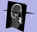
Segmentation of eye ball
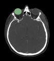
Same segmentation in a 2D view
Key Investigators
- Georgia Tech: Ivan Kolesov and Allen Tannenbaum
- MGH: Gregory Sharp
Objective
- We are interested in segmenting the eye ball, lens, optic nerve, and the optic chiasm.
- Anatomical structures are highly sensitive to radiation.
- We are creating a framework to perform these segmentations, which is likely to require a different approach for each structures due to the proximity of multiple structures to each other.
Approach
- Pivotal organ is considered the eye since its segmentation will localize the region of interest when looking for other structures.
- We will reduce the dimensionality of this problem by performing model based segmentation for each structure.
- Once the eye is segmented, we use this knowledge to locate the lens/ initialize optic nerve segmentation.
- We would like to leverage the information provided by a CT scan with additional data from an MRI -- we have to consider this registration problem.
Progress
- We have the eye ball segmentation.