2011 Winter Project Week:longitudinal dti analysis
Longitudinal DTI Analysis for patient follow-up images- complete processing pipeline


Key Partners
- Utah: Anuja Sharma, Guido Gerig
- UNC: Martin Styner (Core 1)
- Iowa: Hans Johnson (HD Project)
- UCLA: Jack Van Horn (TBI Project)
Objective
To work on longitudinal DTI data from Traumatic Brain Injury data sets and Huntington's disease datasets. The aim is to analyze changes in diffusion in individual patients' follow up images. In the process, explore the inventory of tools needed (existing within or outside Slicer/ITK) and challenges faced in achieving the same, focusing mainly on DTI registration.
Approach, Plan
Begin with utilizing the existing DTI registration resources for co-registering the images from individual subjects (at varying timepoints: intra-subject). The input images would be scalars derived from the DWI/DTI inputs. For the specific scenarios of TBI and HD, different algorithms/parameter settings for registration would be compared. The aim is to build an atlas using the transformed images and getting transformation fields back to each timepoint image. This would be applied to deform the tensor fields and finally come up with a DTI atlas.
Post successful DTI atlas building, we would proceed with tractography in the DTI atlas image, transformed back again to get the same tract geometry from individual images. This is followed by arc-length parametrization along the fiber bundles and the use of existing DTI-statistical-analysis framework with along-tract kernel regression. The framework was originally developed by Casey Goodlett and has been modified and updated by the Utah and UNC groups.
Progress
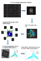
We began with exploring the non rigid Group-wise registration tool using the B-Spline deformation model (Golland et al.) available within the NAMIC kit to come up with a DTI Atlas. We explored a variety of options within it including different levels of multi-resolution optimization, number of iterations and B-Spline grid sizes. The affine step was not working as expected probably because of some further tuning needed within the parameters. So we tried to use the results of manual affine registration using Slicer as a prior to the groupwise tool. We still have to tweak the parameters around to get fine results for the mean atlas and the transformation fields.
As another alternative, we are exploring the use of the DTI registration capability of Slicer. We found that once we get the transformed DTI images, we have tools to get the average DTI atlas for doing tractography. The required piece of interface here is a tool to use the deformation fields (corresponding to each transform between the individual image and the atlas) to map the tract back to individual subjects. Once we have the along tract information from each timepoint, we can use the DTI Tract Statistics tool to further statistically explore the localized variation of diffusion due to pathology or injury. Similarly, if we register the scalar images using Slicer, we need a tool which would provide the deformation fields (forward and backward mapping between individual subjects and atlas). The remaining steps could be done using tools developed by Casey Goodlet et al.
Also, as a part of discussion with the UNC and the Iowa group, we discussed possible changes and improvements needed in the DTI tract statistics command line module developed and tested last year during AHM 2010.
References
- Casey B. Goodlett, P. Thomas Fletcher, John H. Gilmore, Guido Gerig. Group Analysis of DTI Fiber Tract Statistics with Application to Neurodevelopment. NeuroImage 45 (1) Supp. 1, 2009. p. S133-S142