2012 Summer Project Week:4D shape analysis
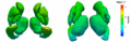
Shape evolution of sub-cortical structures. Color denotes magnitude of velocity.

Key Investigators
- Utah: James Fishbaugh, Marcel Prastawa, Guido Gerig
Objective
We are interested in detecting and quantifying shape changes associated with the progression of Huntington's disease. Preliminary experiments on sub-cortical structures in HD from previous project weeks were intended as a test of our methodology. In order to have an impact moving forward, our tools needs to be robust, easy to use, and available as open-source software. The objective is to improve upon our research code base, leveraging open source libraries.
Approach, Plan
We will consider various software design decisions and begin improving research code.
Progress
We met with current and prospective users to get their input on what they expect from the software.
We considered the input format, which presents a trade off between ease of use and robustness. The easiest input format from a user's point of view would be segmentation label images. However, this choice leads to issues with mesh extraction, simplification, smoothing, etc. We have concluded that the most realistic input to the system should be aligned, simplified meshes. It is up to the user to provide aligned meshes of adequate quality and reasonable complexity. However, the users have requested documentation for the preprocessing steps, with specific tools provided for each step.
We considered the output format as well. We have decided on outputting shapes and corresponding vector fields in ascii VTK format.
A page for downloading the software and documentation for building/using it can be found http://www.cs.utah.edu/~jfishbau/shapereg.html. The page is not yet complete, and the code is not yet available. It will be available soon and this wiki will be updated.
The software was used successfully by Beatriz Paniagua to estimate a healthy atlas from over 400 shapes. The results can be seen below.


References
- Fishbaugh, J., Prastawa, M., Durrleman, S., Piven, J., Gerig G. Analysis of Longitudinal Shape Variability via Subject Specific Growth Modeling. Proc. of Medical Image Computing and Computer Assisted Intervention (MICCAI '12). (to appear),
- Fishbaugh, J., Durrleman, S., Piven, J., Gerig, G. A Framework for Longitudinal Data Analysis via Shape Regression . SPIE Medical Imaging 2012: Image Processing. Vol. 8314.
- Fishbaugh, J., Durrleman, S., Gerig, G. Estimation of Smooth Growth Trajectories with Controlled Acceleration from Time Series Shape Data. Proc. of Medical Image Computing and Computer Assisted Intervention (MICCAI '11).