Difference between revisions of "2013 Summer Project Week:3D prostate segmentation of Ultrasound image"
From NAMIC Wiki
| Line 27: | Line 27: | ||
<div style="width: 27%; float: left; padding-right: 3%;"> | <div style="width: 27%; float: left; padding-right: 3%;"> | ||
<h3>Approach, Plan</h3> | <h3>Approach, Plan</h3> | ||
| − | + | Our proach is localization active contour, we use the ball which centred at the coutour to make the chan-vese method more localization. | |
</div> | </div> | ||
Revision as of 17:10, 17 June 2013
Home < 2013 Summer Project Week:3D prostate segmentation of Ultrasound image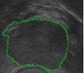
The segmentation result of localized active contour.


Key Investigators
- Nanjing University of Science and Technology: Xu Li
- BWH: Andriy Fedorov, Tina Kapur , William Wells
Objective
Segmentation of prostate gland from 3D Transrectal Ultrasound images. Our goal is to work out a deformable model that can segment the prostate gland well, we are trying to find an appropriate energy function that can have good segmentation result for Ultrasound images.
Approach, Plan
Our proach is localization active contour, we use the ball which centred at the coutour to make the chan-vese method more localization.
Progress
Delivery Mechanism
This work is in very early stages of exploration.
References
JieHuang,Xiaoping Yang,Yunmei Chen. A fast algorithm for global minimization of maximum liklihood based on ultrasound image segmentation. Inverse Problems and Imaging.Volume 5, No. 3, 2011, 645–657.