Difference between revisions of "2014 Project Week:CardiacStemCellMonitoring"
From NAMIC Wiki
Ktdiedrich (talk | contribs) |
Ktdiedrich (talk | contribs) |
||
| Line 37: | Line 37: | ||
<ul> | <ul> | ||
<li>Split 3 plane MRI locator images into separate planes to use axial slices for image registration</li> | <li>Split 3 plane MRI locator images into separate planes to use axial slices for image registration</li> | ||
| + | <li>Enlarged the moving image with a 7 degrees of free transformation </li> | ||
| + | <ul> | ||
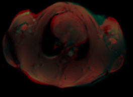
| + | <li>Rigid 6 degree [[File:MovedLinear1fusion.png]]</li> | ||
| + | <li>With Scale 7 degree [[File:MovedScaled1fusion.png]]</li> | ||
| + | </ul> | ||
</ul> | </ul> | ||
</div> | </div> | ||
</div> | </div> | ||
Revision as of 21:41, 8 January 2014
Home < 2014 Project Week:CardiacStemCellMonitoringProject Description
Monitoring engrafted stem cells in cardiac tissue with time series manganese enhanced MRI
Objective
- The goal of the entire project is to restore cardiac function after a heart attack by injecting cardiac stem cells in the infarcted tissue.
- This part of the project seeks to monitor injected stem cells over time.
Approach, Plan
- Pigs (and soon mice) are given a heart attack by inflating a balloon in an artery
- Transgenic stem cells over expressing manganese importer are injected into the infarcted cardiac tissue
- Images are taken of the hearts at stem cell engraftment day 0, 7, and 45
- The images taken are
- PET-CT: PET signal is expected to be reduced in the infarcted tissue ROI on day 0 and increase over day 7 and day 45 as stem cells grow.
- Post GD: Gadolinium Enhanced MRI signal is expected to be high in the infarcted tissue ROI on day 0 and decrease over day 7 and day 45 as stem cells grow and replace the dead tissue.
- Post MN: Manganese is added a contrast agent, the signal is expected to be low in the infarcted tissue ROI on day 0 and increase over day 7 and day 45 as stem cells grow.
- The pigs are growing over the time points by as much as 30% over day 0 to 45.
- Register the locator MRIs of the 3 time points together and apply the transformations to to Post MN images. The Post MN images are too narrow to register directly but were taken in the same frame of reference with low resolution locator or scout images. The registration needs to scale based on the change in heart volume. The change in heart volume may be less than the change in total volume of the pigs.
- Register The time points by CT and then apply the transformations to the PET.
- Copy an ROI across the time points to measure the change in Post MN MRI, Post GD MRI, and PET signal over day 0, 7, 45
Progress
- Split 3 plane MRI locator images into separate planes to use axial slices for image registration
- Enlarged the moving image with a 7 degrees of free transformation
- Rigid 6 degree
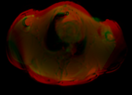
- With Scale 7 degree