2014 Project Week:Multi-Tissue Stroke Segmentation
From NAMIC Wiki
Revision as of 19:16, 17 December 2013 by Rameshvs (talk | contribs) (Created page with '__NOTOC__ <gallery> Image:PW-SLC2014.png|Projects List Image:WMH_T1.png| T1 images in stroke dataset. Image:WMHseg.png | left: FLAIR ima…')
Home < 2014 Project Week:Multi-Tissue Stroke Segmentation
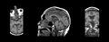
T1 images in stroke dataset.
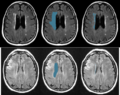
left: FLAIR images, middle: manual delineation of relevant areas, right: manual WMH segmentation.


Key Investigators
- Ramesh Sridharan, Adrian Dalca, Polina Binder, Polina Golland, MIT
- Natalia Rost, Jonathan Rosand, MGH
Project Description
Objective
We have developed some methods for segmentation of white matter hyperintensity (WMH) in FLAIR images of stroke patients. We want to extend our framework to do multi-modal segmentation of multiple tissue types (in our case, stroke lesions, white matter hyperintensity, and normal tissue using T1, FLAIR, DWI, and possibly ADC images). This dataset is particularly challenging due to the low resolution (typically 1mm x 1mm x 7mm) and cropped fields of view in the given images.
Approach, Plan
- Identify intensity and shape signatures of different tissue types across images