Difference between revisions of "2015 Multimodal Guidance for Breast Cancer Surgery"
From NAMIC Wiki
| (7 intermediate revisions by 2 users not shown) | |||
| Line 2: | Line 2: | ||
<gallery> | <gallery> | ||
Image:PW-Summer2015.png|[[2015_Summer_Project_Week#Projects|Projects List]] | Image:PW-Summer2015.png|[[2015_Summer_Project_Week#Projects|Projects List]] | ||
| + | Image:Mikael-Capture2.PNG|Fig 1. 'Stage 1' | ||
| + | Image:Mikael-Capture3.PNG|Fig 2. 'Stage 2' | ||
| + | Image:Mikael-Capture4.PNG|Fig 3. Hitachi US | ||
| + | Image:Mikael-Capture5.PNG|Fig 4. Philips US | ||
| + | Image:Mikael-Capture1.PNG|Fig 5. 3D Slicer | ||
</gallery> | </gallery> | ||
==Key Investigators== | ==Key Investigators== | ||
| − | * Mikael Brudfors | + | * Mikael Brudfors, Universidad Carlos III de Madrid (UC3M) --- formerly KTH & UBC, next UCL |
| − | * David García | + | * David García, UC3M |
| − | * Javier Pascau ( | + | * Javier Pascau, UC3M |
| + | * Tamas Ungi, Andras Lasso (Queen's University, Kingsotn, ON, Canada) | ||
==Project Description== | ==Project Description== | ||
| Line 13: | Line 19: | ||
<div style="width: 27%; float: left; padding-right: 3%;"> | <div style="width: 27%; float: left; padding-right: 3%;"> | ||
<h3>Objective</h3> | <h3>Objective</h3> | ||
| − | * Development of an intraoperative guidance system for | + | * Development of an intraoperative guidance system for breast cancer surgery that would allow for guidance, tumor detection and localization. |
</div> | </div> | ||
<div style="width: 27%; float: left; padding-right: 3%;"> | <div style="width: 27%; float: left; padding-right: 3%;"> | ||
<h3>Approach, Plan</h3> | <h3>Approach, Plan</h3> | ||
| − | * | + | * Possibilities of combining multiple imaging modalities (3D Scanner Surface data, US, MR) and visualizing these in 3D Slicer |
</div> | </div> | ||
<div style="width: 27%; float: left; padding-right: 3%;"> | <div style="width: 27%; float: left; padding-right: 3%;"> | ||
| Line 24: | Line 30: | ||
* Record surface data using an Artec 3D scanner and visualize it in 3D Slicer (DONE) | * Record surface data using an Artec 3D scanner and visualize it in 3D Slicer (DONE) | ||
* Communicate with the 3D Scanner through 3D slicer module (WORK IN PROGRESS) | * Communicate with the 3D Scanner through 3D slicer module (WORK IN PROGRESS) | ||
| + | * Define an acquisition protocol. Tamas Ungi showed their protocol and Slicer module that includes needle tracking and a possible collaboration was identified (WORK IN PROGRESS) | ||
</div> | </div> | ||
</div> | </div> | ||
Latest revision as of 15:20, 24 June 2015
Home < 2015 Multimodal Guidance for Breast Cancer Surgery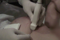
Fig 1. 'Stage 1'
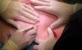
Fig 2. 'Stage 2'
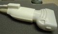
Fig 3. Hitachi US
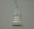
Fig 4. Philips US
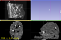
Fig 5. 3D Slicer


Key Investigators
- Mikael Brudfors, Universidad Carlos III de Madrid (UC3M) --- formerly KTH & UBC, next UCL
- David García, UC3M
- Javier Pascau, UC3M
- Tamas Ungi, Andras Lasso (Queen's University, Kingsotn, ON, Canada)
Project Description
Objective
- Development of an intraoperative guidance system for breast cancer surgery that would allow for guidance, tumor detection and localization.
Approach, Plan
- Possibilities of combining multiple imaging modalities (3D Scanner Surface data, US, MR) and visualizing these in 3D Slicer
Progress
- Acquire tracked US using the PLUS toolkit (DONE)
- Record surface data using an Artec 3D scanner and visualize it in 3D Slicer (DONE)
- Communicate with the 3D Scanner through 3D slicer module (WORK IN PROGRESS)
- Define an acquisition protocol. Tamas Ungi showed their protocol and Slicer module that includes needle tracking and a possible collaboration was identified (WORK IN PROGRESS)