Difference between revisions of "2017 Winter Project Week/CoronarySegmentationTool"
From NAMIC Wiki
Zhouhaoyin (talk | contribs) m |
Zhouhaoyin (talk | contribs) |
||
| Line 3: | Line 3: | ||
Image:PW-Winter2017.png|link=2017_Winter_Project_Week#Projects|[[2017_Winter_Project_Week#Projects|Projects List]] | Image:PW-Winter2017.png|link=2017_Winter_Project_Week#Projects|[[2017_Winter_Project_Week#Projects|Projects List]] | ||
<!-- Use the "Upload file" link on the left and then add a line to this list like "File:MyAlgorithmScreenshot.png" --> | <!-- Use the "Upload file" link on the left and then add a line to this list like "File:MyAlgorithmScreenshot.png" --> | ||
| + | File:Slicerscreenshot.png|Selecting a vessel segment, new views will be generated and users can manual editing the segmentation result. | ||
</gallery> | </gallery> | ||
| Line 26: | Line 27: | ||
| | | | ||
<!-- Progress and Next steps bullet points (fill out at the end of project week) --> | <!-- Progress and Next steps bullet points (fill out at the end of project week) --> | ||
| − | * | + | *We applied curved reformation algorithms to provide more intuitive and more user-friendly interfaces. |
|} | |} | ||
Revision as of 18:51, 12 January 2017
Home < 2017 Winter Project Week < CoronarySegmentationTool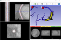
Selecting a vessel segment, new views will be generated and users can manual editing the segmentation result.

Key Investigators
- Haoyin Zhou, Brigham and Women's Hospital
- Jayender Jagadeesan, Brigham and Women's Hospital
Project Description
| Objective | Approach and Plan | Progress and Next Steps |
|---|---|---|
|
|
|