Difference between revisions of "2017 Winter Project Week/Evaluate Deep Learning for binary cancer legion classification"
From NAMIC Wiki
| (16 intermediate revisions by the same user not shown) | |||
| Line 2: | Line 2: | ||
Image:PW-Winter2017.png|link=2017_Winter_Project_Week#Projects|[[2017_Winter_Project_Week#Projects|Projects List]] | Image:PW-Winter2017.png|link=2017_Winter_Project_Week#Projects|[[2017_Winter_Project_Week#Projects|Projects List]] | ||
Image:Cancer_roi_Img_00001.png|Example Cancer ROI | Image:Cancer_roi_Img_00001.png|Example Cancer ROI | ||
| + | Image:Cancer-roi-digits-training-0109.png|Initial LeNet training with DIGITS/Caffe | ||
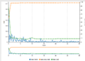
| + | Image:KVis-trained-LeNet-data-augmentation.png|Training performance after data augmentation | ||
| + | KVis-deep-learning-project.pdf | Presentation Slides on the project | ||
| + | KVis-deep-learning-video.mov | Project Demonstration Video | ||
<!-- Use the "Upload file" link on the left and then add a line to this list like "File:MyAlgorithmScreenshot.png" --> | <!-- Use the "Upload file" link on the left and then add a line to this list like "File:MyAlgorithmScreenshot.png" --> | ||
</gallery> | </gallery> | ||
| Line 7: | Line 11: | ||
==Key Investigators== | ==Key Investigators== | ||
<!-- Add a bulleted list of investigators and their institutions here --> | <!-- Add a bulleted list of investigators and their institutions here --> | ||
| − | Curt Lisle, KnowledgeVis, LLC | + | *Curt Lisle, KnowledgeVis, LLC |
| − | + | *Yanling Liu, FNLCR | |
==Project Description== | ==Project Description== | ||
| Line 21: | Line 25: | ||
| | | | ||
<!-- Approach and Plan bullet points --> | <!-- Approach and Plan bullet points --> | ||
| − | * | + | * Start with a dataset, prepared at the Frederick National Lab for Cancer Research, to use to train a classifier. |
| − | * The | + | * The dataset consists of a series of 52x52 Region Of Interest T2 MR images containing cancer lesions and two T2 image series which do not contain lesions. |
| − | * We plan to collect advice from others at the Project Week | + | * We plan to collect advice from others at the Project Week, select a deep learning framework, and attempt to build a classifer using this training data. |
| | | | ||
<!-- Progress and Next steps bullet points (fill out at the end of project week) --> | <!-- Progress and Next steps bullet points (fill out at the end of project week) --> | ||
| − | * | + | * created a DIGITS Amazon instance using NVIDIA's marketplace image before project week |
| + | * Prepared the dataset in the style of the MNIST example | ||
| + | * Trained LeNet and AlexNet CNNs using DIGITS interface and Caffe learning framework | ||
| + | * Data augmentation was crucial to improve results up to 83% detection accuracy for 2D case | ||
| + | * 3D data was presented without augmentation and yielded better results than 2D alone | ||
| + | * We believe results will further improve when better data augmentation and 3D slice data are used simultaneously | ||
|} | |} | ||
==Background and References== | ==Background and References== | ||
<!-- Use this space for information that may help people better understand your project, like links to papers, source code, or data --> | <!-- Use this space for information that may help people better understand your project, like links to papers, source code, or data --> | ||
Latest revision as of 15:35, 13 January 2017
Home < 2017 Winter Project Week < Evaluate Deep Learning for binary cancer legion classification
Example Cancer ROI
Initial LeNet training with DIGITS/Caffe

Training performance after data augmentation

Presentation Slides on the project
Project Demonstration Video


Key Investigators
- Curt Lisle, KnowledgeVis, LLC
- Yanling Liu, FNLCR
Project Description
| Objective | Approach and Plan | Progress and Next Steps |
|---|---|---|
|
|
|