Difference between revisions of "2017 Winter Project Week/MeningiomaSegmentation"
From NAMIC Wiki
(Created page) |
(added progress and next steps) |
||
| Line 3: | Line 3: | ||
Image:PW-Winter2017.png|link=2017_Winter_Project_Week#Projects|[[2017_Winter_Project_Week#Projects|Projects List]] | Image:PW-Winter2017.png|link=2017_Winter_Project_Week#Projects|[[2017_Winter_Project_Week#Projects|Projects List]] | ||
<!-- Use the "Upload file" link on the left and then add a line to this list like "File:MyAlgorithmScreenshot.png" --> | <!-- Use the "Upload file" link on the left and then add a line to this list like "File:MyAlgorithmScreenshot.png" --> | ||
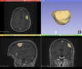
| + | Image:Jk_meningioma_seg.png|link=File:Jk_meningioma_seg.png|[[Jk_meningioma_seg.png|Meningioma Segmentation]] | ||
</gallery> | </gallery> | ||
| Line 25: | Line 26: | ||
| | | | ||
<!-- Progress and Next steps bullet points (fill out at the end of project week) --> | <!-- Progress and Next steps bullet points (fill out at the end of project week) --> | ||
| − | * | + | Progress |
| + | * Segmented with ANTs and FSL. | ||
| + | * Learned about Slicer segmentation tools, and segmented semi-automatically. | ||
| + | * Put in contact with people who have segmented meningiomas. | ||
| + | |||
| + | Next steps | ||
| + | * Continue learning about what has been done in the past. | ||
| + | * Improve brain-extraction. | ||
| + | * Continue testing existing segmentation methods. | ||
| + | * Try Slicer in a Nipype workflow. | ||
| + | * Apply manifold learning (a method of dimensionality reduction). | ||
|} | |} | ||
Revision as of 01:11, 13 January 2017
Home < 2017 Winter Project Week < MeningiomaSegmentationKey Investigators
- Satrajit Ghosh, MIT
- Omar Arnaout, Brigham and Women's Hospital
Project Description
| Objective | Approach and Plan | Progress and Next Steps |
|---|---|---|
|
|
Progress
Next steps
|
Background and References
MR images of meningiomas that will be used in this project are available at OpenNeu.ro.

