Difference between revisions of "2017 Winter Project Week/Multi-ModalitySegmentationOfUSandMRImagesForGliomaSurgery"
From NAMIC Wiki
(Created page with "__NOTOC__ <gallery> Image:PW-Winter2017.png|link=2017_Winter_Project_Week#Projects|Projects List <!-- Use the "Upload file" link on the l...") |
|||
| Line 3: | Line 3: | ||
Image:PW-Winter2017.png|link=2017_Winter_Project_Week#Projects|[[2017_Winter_Project_Week#Projects|Projects List]] | Image:PW-Winter2017.png|link=2017_Winter_Project_Week#Projects|[[2017_Winter_Project_Week#Projects|Projects List]] | ||
<!-- Use the "Upload file" link on the left and then add a line to this list like "File:MyAlgorithmScreenshot.png" --> | <!-- Use the "Upload file" link on the left and then add a line to this list like "File:MyAlgorithmScreenshot.png" --> | ||
| + | File:USandMRIimagesMLSegmentation.png|Several structures of the brain segmented from US- and MR images. | ||
</gallery> | </gallery> | ||
Revision as of 16:42, 9 January 2017
Home < 2017 Winter Project Week < Multi-ModalitySegmentationOfUSandMRImagesForGliomaSurgery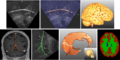
Several structures of the brain segmented from US- and MR images.

Key Investigators
- Jennifer Nitsch, University of Bremen (Germany)
Project Description
| Objective | Approach and Plan | Progress and Next Steps |
|---|---|---|
|
|
|