Difference between revisions of "2017 Winter Project Week/Multi-ModalitySegmentationOfUSandMRImagesForGliomaSurgery"
From NAMIC Wiki
| (4 intermediate revisions by the same user not shown) | |||
| Line 4: | Line 4: | ||
<!-- Use the "Upload file" link on the left and then add a line to this list like "File:MyAlgorithmScreenshot.png" --> | <!-- Use the "Upload file" link on the left and then add a line to this list like "File:MyAlgorithmScreenshot.png" --> | ||
File:USandMRIimagesMLSegmentation.png|Several structures of the brain segmented from US- and MR images. | File:USandMRIimagesMLSegmentation.png|Several structures of the brain segmented from US- and MR images. | ||
| + | File:Dnn.png | ||
| + | File:SkullStrip.tcsproj.mp4 | ||
</gallery> | </gallery> | ||
| Line 9: | Line 11: | ||
<!-- Add a bulleted list of investigators and their institutions here --> | <!-- Add a bulleted list of investigators and their institutions here --> | ||
* Jennifer Nitsch, University of Bremen (Germany) | * Jennifer Nitsch, University of Bremen (Germany) | ||
| + | * Hans Meine, University of Bremen (Germany) | ||
| Line 19: | Line 22: | ||
| | | | ||
<!-- Objective bullet points --> | <!-- Objective bullet points --> | ||
| − | * Multi-Modual Image Segmentation of preoperative MR- and intraoperative Ultrasound(US)-images | + | * Multi-Modual Image Segmentation of preoperative MR- and intraoperative Ultrasound(US)-images for multi-modual image registration. |
* Segmentation of the following anatomical structures: Falx cerebri, tentorium cerebelli, white matter, gray matter, CSF (ventricles), blood vessels. | * Segmentation of the following anatomical structures: Falx cerebri, tentorium cerebelli, white matter, gray matter, CSF (ventricles), blood vessels. | ||
* Testing applicability of Deep Learing on current data | * Testing applicability of Deep Learing on current data | ||
| Line 29: | Line 32: | ||
| | | | ||
<!-- Progress and Next steps bullet points (fill out at the end of project week) --> | <!-- Progress and Next steps bullet points (fill out at the end of project week) --> | ||
| − | * | + | *Trained a DNN for skull stripping on 18 data sets from a public database, also segmented in GM, WM, CSF |
| + | *There a a lot of databases for T1-weighted brain images, but good reference data is rare... | ||
|} | |} | ||
==Background and References== | ==Background and References== | ||
<!-- Use this space for information that may help people better understand your project, like links to papers, source code, or data --> | <!-- Use this space for information that may help people better understand your project, like links to papers, source code, or data --> | ||
Latest revision as of 14:37, 16 January 2017
Home < 2017 Winter Project Week < Multi-ModalitySegmentationOfUSandMRImagesForGliomaSurgery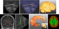
Several structures of the brain segmented from US- and MR images.


Key Investigators
- Jennifer Nitsch, University of Bremen (Germany)
- Hans Meine, University of Bremen (Germany)
Project Description
| Objective | Approach and Plan | Progress and Next Steps |
|---|---|---|
|
|
|