Difference between revisions of "Analysis of different atlas-based segmentation techniques for parotid glands"
Kfritscher76 (talk | contribs) |
Kfritscher76 (talk | contribs) |
||
| Line 31: | Line 31: | ||
3 different segmentation approaches based on the usage of multiple atlases [1], statistical appearance models [2] and a method based on using image patches in combination with Gaussian processes for segmentation have been tested for their suitability to segment the parotid gland using a dataset of 18 CT images. Different approaches to combine the atlas and model based approaches [1,2] in different ways are currently under development. | 3 different segmentation approaches based on the usage of multiple atlases [1], statistical appearance models [2] and a method based on using image patches in combination with Gaussian processes for segmentation have been tested for their suitability to segment the parotid gland using a dataset of 18 CT images. Different approaches to combine the atlas and model based approaches [1,2] in different ways are currently under development. | ||
| − | |||
| − | |||
| − | |||
| − | |||
| − | |||
| + | '''Methods:<br>''' | ||
| − | First tests using following pipelines: | + | *LF = Image-based Registration with Label Fusion |
| − | PatchClass -> SAM | + | *SAM = Statistical Appearance Models (label map registration, PCA, etc) |
| − | LF -> GP | + | *PatchClass = Patch classification with Random Forest |
| − | + | *GP = Gaussian Processes for label refinement | |
| − | PatchClass -> SAM -> GP | + | |
| − | LF -> GP -> SAM | + | <br>20 test datasets (CTs) of Head-Neck region have been exchanged |
| + | |||
| + | |||
| + | '''First tests using following pipelines will be carried out:''' | ||
| + | *PatchClass -> SAM<br> | ||
| + | *LF -> GP<br> | ||
| + | ''' | ||
| + | '''Further potential combinations:<br>''' | ||
| + | *PatchClass -> SAM -> GP | ||
| + | *LF -> GP -> SAM | ||
</div> | </div> | ||
Revision as of 13:05, 21 June 2013
Home < Analysis of different atlas-based segmentation techniques for parotid glands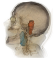
Parotid gland + brainstem


Key Investigators
- MIT: Christian Wachinger, Matthew Brennan
- MGH: Karl Fritscher, Greg Sharp
Objective
Our goal is to investigate various segmentation approaches for identifying parotid glands on head and neck CT images. The focus will be on atlas-based methods, which exploit the information from a number of previously labeled images. Several different strategies exist on how to employ this prior information to achieve the segmentation.
Plan
We will try to work out the differences in terms of parameterization and regularization of various atlas-based methods. We will further try to characterize properties of such methods for the segmentation of parotid glands, which show high structural variability. Finally, we would like to investigate, which combination of methods may be promising.
Progress
3 different segmentation approaches based on the usage of multiple atlases [1], statistical appearance models [2] and a method based on using image patches in combination with Gaussian processes for segmentation have been tested for their suitability to segment the parotid gland using a dataset of 18 CT images. Different approaches to combine the atlas and model based approaches [1,2] in different ways are currently under development.
Methods:
- LF = Image-based Registration with Label Fusion
- SAM = Statistical Appearance Models (label map registration, PCA, etc)
- PatchClass = Patch classification with Random Forest
- GP = Gaussian Processes for label refinement
20 test datasets (CTs) of Head-Neck region have been exchanged
First tests using following pipelines will be carried out:
- PatchClass -> SAM
- LF -> GP
Further potential combinations:
- PatchClass -> SAM -> GP
- LF -> GP -> SAM
References
- [1] Peroni M, Methods and Algorithms for Image Guided Adaptive Radio- and Hadron Therapy. PhD Thesis, Politecnico di Milano, 2011
- [2] Fritscher KD, Gruenerbl A, Schubert R, 3D image segmentation using combined shape-intensity prior models. Journal of Computer Assisted Radiology and Surgery, 2007;1:341–350
- [3] Wachinger C, Sharp G, Golland P, Contour-Driven Regression for Label Inference in Atlas-Based Segmentation, MICCAI, 2013.