Clinically oriented assessment of local changes in the properties of white matter affected by intra-cranial hemorrhage
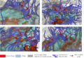
Peri-lesional deformation / bending of fiber tracts around pathology
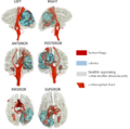
Identification of corticospinal tract in patient with acute TBI and intracranial hemorrhage
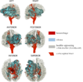
Challenges of identifying corticospinal tract in the presence of large isotropy in hemorrhagic regions


Key Investigators
- UCLA: Andrei Irimia, Micah Chambers, Ron Kikinis, Jack Van Horn
Objective
Intra-cranial hemorrhage (ICH) is a phenomenon which is frequently associated with traumatic brain injury (TBI) and which can be life-threatening depending on the location, extent and overall severity of the bleeding. Extensive ICH is associated with clinical outcome which is poorer than in cases where this phenomenon is not detectable via GRE T2 or SWI imaging. Identification of ICH is important in a clinical setting because it can indicate TBI severity and also provide useful information to neurosurgeons in the critical hours and days following TBI. Of particular interest to TBI-related ICH researchers is the ability to assess local changes in the properties of white matter (WM), particularly at the boundary between contusional and healthy-appearing tissues, known as the peri-hemorrhagic regions. The purpose of this project is to use 3D Slicer to identify and characterize the putative boundary between ICH and healthy-appearing tissues.
Approach, Plan
- We seek to use a multimodal imaging approach (T1/T2, FLAIR and SWI imaging) to identify the peri-lesional and especially peri-hemorrhagic regions of the brain in several test cases
- Tissue classification and model rendering will be performed within 3D Slicer using existing tools to distinguish between hemorrhage and health-appearing WM
- We are interested in using 3D Slicer to perform a rigorous quantification of what happens to peri-hemorrhagic fiber bundles as a result of extensive bleeding in the brain
Progress
References
- Andrei Irimia and John D. van Horn (2012) The structural, connectomic and network covariance of the human brain NeuroImage (accepted) [1]
- Andrei Irimia, Bo Wang, Stephen R. Aylward, Marcel W. Prastawa, Danielle F. Pace, Marc Niethammer, Guido Gerig, David A. Hovda, Ron Kikinis, Paul M. Vespa and John D. van Horn (2012) Multimodal neuroimaging of structural pathology and connectomics in traumatic brain injury: toward personalized outcome prediction Neuroimage: Clinical volume 1, pages 1-17 [2]
- John D. van Horn, Andrei Irimia, Carinna M. Torgerson, Micah C. Chambers, Arthur W. Toga (2012) Mapping connectivity damage in the case of Phineas Gage. Plos ONE [3]
- Andrei Irimia, Micah C. Chambers, Carinna M. Torgerson and John D. van Horn (2012) Circular representation of human cortical networks for subject and population-level connectomic visualization NeuroImage volume 60, pages 1340-1351
- Andrei Irimia, Micah C Chambers, Carinna M Torgerson, Maria Filippou, David A Hovda, Jeffry R Alger, Guido Gerig, Arthur W Toga, Paul M Vespa, Ron Kikinis, John D Van Horn (2012) Patient-tailored connectomics visualization for the assessment of white matter atrophy in traumatic brain injury. Frontiers in Neurology [4]
- Andrei Irimia, Micah C. Chambers, Jeffry R. Alger, Maria Filippou, Marcel W. Prastawa, Bo Wang, David A. Hovda, Guido Gerig, Arthur W. Toga, Ron Kikinis, Paul M. Vespa, John D. van Horn (2011) Comparison of acute and chronic traumatic brain injury using semi-automatic multimodal segmentation of MR volumes. Journal of Neurotrauma [5]