Collaboration/UIowa/Developing a GUI for non-rigid image registration programs using NA-MIC Kit
 Return to Project Week Main Page |
Key Investigators
- Gary Christensen, The University of Iowa
- Kunlin Cao, The University of Iowa
- Kai Ding, The University of Iowa
- Xiujuan Geng, The University of Iowa
- Paul Song, The University of Iowa
- Hans Jonson, The University of Iowa
- Yumin, Kitware
Objective
Learn how to use a professional software development process. Learn how to create GUIs to make our tools easier for others to use. Learn how to port our existing image registration & image processing libraries and tools to the ITK and VTK model. Learn how to package our programs so they run on different computer operating systems such as Windows, Linux, and Macintosh.
Approach, Plan
Our plan for the project week is to
- Learn about the image processing pipeline and memory management issues.
- Learn about ITK file formats.
- Learn how to read, write, and visualize images using NA-MIC tools.
- Learn how to make GUIs ( Evaluate whether or not we need to write our own GUI using KWWidgets or can take advantage of slicer3 )
- Develop an Analyze object map reader.
- Learn how to use cpack.
The GUI should have the following components
- Show the template and target images before registration to verify input. Views would include 2D and 3D views. The GUI would also show landmarks, contours, and surfaces to be used for registration and for validation of the input.
- Allow the user to set various parameters. Allow the user to label corresponding landmarks and contours (possibly surfaces in the future) in the images.
- Show status and instant messages while the program is running, i.e., deforming image, difference images, Jacobian, displacement fields, deforming grid, etc.
Progress
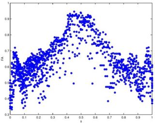
Software for the fiber tracking and statistical analysis along the tracts has been implemented. The statistical methods for diffusion tensors are implemented as ITK code as part of the DTI Software Infrastructure project. The methods have been validated on a repeated scan of a healthy individual. This work has been published as a conference paper (MICCAI 2005) and a journal version (MEDIA 2006). Our recent IPMI 2007 paper includes a nonparametric regression method for analyzing data along a fiber tract.
References
- Fletcher, P.T., Tao, R., Jeong, W.-K., Whitaker, R.T., "A Volumetric Approach to Quantifying Region-to-Region White Matter Connectivity in Diffusion Tensor MRI," to appear Information Processing in Medical Imaging (IPMI) 2007.
- Corouge, I., Fletcher, P.T., Joshi, S., Gilmore, J.H., and Gerig, G., "Fiber Tract-Oriented Statistics for Quantitative Diffusion Tensor MRI Analysis," Medical Image Analysis 10 (2006), 786--798.
- Corouge, I., Fletcher, P.T., Joshi, S., Gilmore J.H., and Gerig, G., Fiber Tract-Oriented Statistics for Quantitative Diffusion Tensor MRI Analysis, Lecture Notes in Computer Science LNCS, James S. Duncan and Guido Gerig, editors, Springer Verlag, Vol. 3749, Oct. 2005, pp. 131 -- 138
- C. Goodlett, I. Corouge, M. Jomier, and G. Gerig, A Quantitative DTI Fiber Tract Analysis Suite, The Insight Journal, vol. ISC/NAMIC/ MICCAI Workshop on Open-Source Software, 2005, Online publication: http://hdl.handle.net/1926/39 .

