Difference between revisions of "DBP3:Utah:RegSegPipeline"
From NAMIC Wiki
| Line 3: | Line 3: | ||
== Pilot Studies on a Registration & Segmentation Pipeline & Workflow == | == Pilot Studies on a Registration & Segmentation Pipeline & Workflow == | ||
| − | + | Overall processing steps are (order tentative) | |
| − | + | #N4 bias field correction for the MRI (surface coils): | |
| − | + | ## run on entire image gives some benefit that '''may''' be improved with masking: again the dominant intensity dropoff from the surface coil occurs along the chest wall and ribcage. Even if that is not the structure of interest, it is the low-freq. variation the bias correction algorithm is searching for, and masking that out can be counter-productive: via masking we may end up with a smoother image, but the intensity variations removed were not caused by the coil but are actually true signal. | |
| − | + | ##Module used: [http://www.slicer.org/slicerWiki/index.php/Modules:N4ITKBiasFieldCorrection-Documentation-3.6 N4 ITK] | |
| − | + | #registration MRA>cMRI | |
| − | + | ##the MRA contains the same FOV and has surrounding structures (liver, chest, spine etc) visible also, despite lower intensities. A global affine is thus not necessarily going to benefit from masking the heart, unless the relative motion of the heart becomes the dominant reason for misalignment. | |
| − | + | ##Module used: [http://www.slicer.org/slicerWiki/index.php/ModulesBRAINSfit-Documentation-3.6 BRAINSfit] | |
| − | + | #ROI definition (manual box ROI or automated via atlas) | |
| − | + | #segmentation of LA from MRA -> inner wall | |
| + | ##as a dynamic image the MRA contains significant spread and likely requires interactive segmentation/thresholding to yield a satisfactory LA volume | ||
| + | ##Module used: [http://www.slicer.org/slicerWiki/index.php/Editor-Documentation-3.6 Editor: thresholding] or thresholding within [http://www.slicer.org/slicerWiki/index.php/Volumes-Documentation-3.6 Volumes thresholding option within ''Display'' tab, use ''iron'' colormap & low alpha setting to check for ventricular wall borders.] | ||
| + | |||
| + | ##cropping and island removal | ||
| + | #LA wall segmentation | ||
| + | ## very small structure, most reliably done manually direct. Starting with automation may yield more effort on post-edits | ||
| + | #segmentation of enhancement within LA wall: intensity statistics. An atlas-based set of intensity distributions may be more meaningful here than a simple Otsu, because both amount and location of enhancement is unknown and can in theory be 0. | ||
| + | #registration follow-up -> baseline | ||
| + | ##most reliably done on the post contrast MRI. | ||
| + | ##DOF of 12 or even low-res BSpline should be ok | ||
| + | |||
<gallery perrow="2" widths="200px"> | <gallery perrow="2" widths="200px"> | ||
Image:DBP3_AFib_LV_overlay_1.jpg| Example contrast MRI with thresholded MRA as color overlay | Image:DBP3_AFib_LV_overlay_1.jpg| Example contrast MRI with thresholded MRA as color overlay | ||
Image:DBP3_AFib_LA_overlay_2.jpg|Example contrast MRI with thresholded MRA as color overlay and areas of enhancement marked | Image:DBP3_AFib_LA_overlay_2.jpg|Example contrast MRI with thresholded MRA as color overlay and areas of enhancement marked | ||
</gallery> | </gallery> | ||
Revision as of 21:39, 10 February 2011
Home < DBP3:Utah:RegSegPipelineback to DBP3 home
The CARMA DBP: MRI-based study and treatment of atrial fibrillation
Pilot Studies on a Registration & Segmentation Pipeline & Workflow
Overall processing steps are (order tentative)
- N4 bias field correction for the MRI (surface coils):
- run on entire image gives some benefit that may be improved with masking: again the dominant intensity dropoff from the surface coil occurs along the chest wall and ribcage. Even if that is not the structure of interest, it is the low-freq. variation the bias correction algorithm is searching for, and masking that out can be counter-productive: via masking we may end up with a smoother image, but the intensity variations removed were not caused by the coil but are actually true signal.
- Module used: N4 ITK
- registration MRA>cMRI
- the MRA contains the same FOV and has surrounding structures (liver, chest, spine etc) visible also, despite lower intensities. A global affine is thus not necessarily going to benefit from masking the heart, unless the relative motion of the heart becomes the dominant reason for misalignment.
- Module used: BRAINSfit
- ROI definition (manual box ROI or automated via atlas)
- segmentation of LA from MRA -> inner wall
- as a dynamic image the MRA contains significant spread and likely requires interactive segmentation/thresholding to yield a satisfactory LA volume
- Module used: Editor: thresholding or thresholding within Volumes thresholding option within Display tab, use iron colormap & low alpha setting to check for ventricular wall borders.
- cropping and island removal
- LA wall segmentation
- very small structure, most reliably done manually direct. Starting with automation may yield more effort on post-edits
- segmentation of enhancement within LA wall: intensity statistics. An atlas-based set of intensity distributions may be more meaningful here than a simple Otsu, because both amount and location of enhancement is unknown and can in theory be 0.
- registration follow-up -> baseline
- most reliably done on the post contrast MRI.
- DOF of 12 or even low-res BSpline should be ok
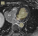
Example contrast MRI with thresholded MRA as color overlay
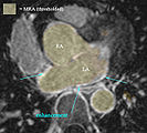
Example contrast MRI with thresholded MRA as color overlay and areas of enhancement marked

