DPB3:Past Featured Articles
From NAMIC Wiki
Home < DPB3:Past Featured Articles
Back to NA-MIC DBP 3
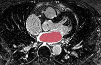
| Atrial fibrillation, a cardiac arrhythmia characterized by unsynchronized electrical activity in the atrial chambers of the heart, is a rapidly growing problem in modern societies. One treatment, referred to as catheter ablation, targets specific parts of the left atrium for radio frequency ablation using an intracardiac catheter. Magnetic resonance imaging has been used for both pre- and and post-ablation assessment of the atrial wall. Magnetic resonance imaging can aid in selecting the right candidate for the ablation procedure and assessing post-ablation scar formations. Image processing techniques can be used for automatic segmentation of the atrial wall, which facilitates an accurate statistical assessment of the region. As a first step towards the general solution to the computer-assisted segmentation of the left atrial wall, in this paper we use shape learning and shape-based image segmentation to identify the endocardial wall of the left atrium in the delayed-enhancement magnetic resonance images. | |
| Gao Y., Gholami B., MacLeod R.S., Blauer J., Haddad W.M., Tannenbaum A. Segmentation of the Endocardial Wall of the Left Atrium using Local Region-Based Active Contours and Statistical Shape Learning. Proceedings of SPIE Medical Imaging 2010. |
