Difference between revisions of "EM Segmentation For Orthopaedic Applications"
From NAMIC Wiki
| Line 6: | Line 6: | ||
* We have utilized the Slicer2.7 EM Segmentation Module for segmentation of the phalanx bones | * We have utilized the Slicer2.7 EM Segmentation Module for segmentation of the phalanx bones | ||
| − | *# Registration was performed outside of | + | *# Registration was performed outside of Slicer using ITK registration algorithms that are available in the IaFeMesh software. This includes Thin plate spline, B-Spline and rigid registration algorithms. |
*# Probability map information was created from a single subject used as the atlas image and filtered using a Gaussian filter. | *# Probability map information was created from a single subject used as the atlas image and filtered using a Gaussian filter. | ||
| − | * Initial evaluation has been | + | * Initial evaluation has been performed |
| + | **Reliability assessed on fourteen specimens that also had the index finger segmented, and two specimens that had the index, middle, ring and little fingers manually segmented | ||
| + | **Validation assessed using the laser scanning of the index finger (proximal, middle, and distal) phalanx bones in five specimens | ||
* Work is underway to develop a Slicer2.7 tutorial for this segmentation ([[Image:Draft_EM_Segment_Tutorial_9_12.pdf|Draft Copy]]) | * Work is underway to develop a Slicer2.7 tutorial for this segmentation ([[Image:Draft_EM_Segment_Tutorial_9_12.pdf|Draft Copy]]) | ||
| Line 21: | Line 23: | ||
'''Links:''' | '''Links:''' | ||
| − | *[http://www.ccad.uiowa.edu/mimx Musculoskeletal Imaging, | + | *[http://www.ccad.uiowa.edu/mimx Musculoskeletal Imaging, Modeling and Experimentation (MIMX)] |
Revision as of 20:10, 6 January 2008
Home < EM Segmentation For Orthopaedic ApplicationsObjective:
- To utilize the Slicer3 Expectation Maximization Algorithm for segmentation of the phalanx bones of the hand.
Progress:
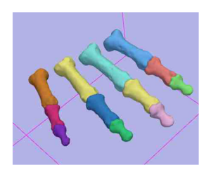
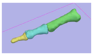
- We have utilized the Slicer2.7 EM Segmentation Module for segmentation of the phalanx bones
- Registration was performed outside of Slicer using ITK registration algorithms that are available in the IaFeMesh software. This includes Thin plate spline, B-Spline and rigid registration algorithms.
- Probability map information was created from a single subject used as the atlas image and filtered using a Gaussian filter.
- Initial evaluation has been performed
- Reliability assessed on fourteen specimens that also had the index finger segmented, and two specimens that had the index, middle, ring and little fingers manually segmented
- Validation assessed using the laser scanning of the index finger (proximal, middle, and distal) phalanx bones in five specimens
- Work is underway to develop a Slicer2.7 tutorial for this segmentation (File:Draft EM Segment Tutorial 9 12.pdf)
To Do:
- Evaluate the Slicer3 EM Segmentation Module
- Update the the tutorial to support the Slicer3 Workflow
- Determine if registration tools in Slicer are adequate for this work.
Key Investigators:
- Iowa: Austin Ramme, Nicole Grosland, and Vincent Magnotta
Links:
Figures:

