Difference between revisions of "File:Histopathology GVD2 grade4 2 zoom.png"
From NAMIC Wiki
(Region segmentation and nuclei detection results for a sample Grade 4 image. Blue: Detected nuclei centers, Red: nuclei boundaries obtained using marker controlled watershed segmentation. Nuclei center recall:81%, precision:96%.) |
(No difference)
|
Latest revision as of 15:15, 20 June 2010
Region segmentation and nuclei detection results for a sample Grade 4 image. Blue: Detected nuclei centers, Red: nuclei boundaries obtained using marker controlled watershed segmentation. Nuclei center recall:81%, precision:96%.
File history
Click on a date/time to view the file as it appeared at that time.
| Date/Time | Thumbnail | Dimensions | User | Comment | |
|---|---|---|---|---|---|
| current | 15:15, 20 June 2010 | 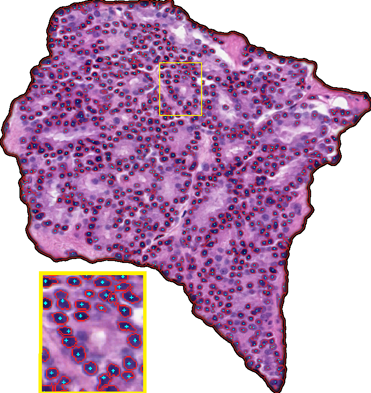 | 762 × 807 (951 KB) | Palani (talk | contribs) | Region segmentation and nuclei detection results for a sample Grade 4 image. Blue: Detected nuclei centers, Red: nuclei boundaries obtained using marker controlled watershed segmentation. Nuclei center recall:81%, precision:96%. |
- You cannot overwrite this file.
File usage
The following page uses this file: