Difference between revisions of "Loading and segmentation of histopathology imaging for radiological-pathological correlation"
From NAMIC Wiki
| Line 8: | Line 8: | ||
==Key Investigators== | ==Key Investigators== | ||
| − | * SPL: Andrey Fedorov | + | * SPL: Tobias Penzkofer, Andrey Fedorov |
<div style="margin: 20px;"> | <div style="margin: 20px;"> | ||
Revision as of 03:18, 20 June 2012
Home < Loading and segmentation of histopathology imaging for radiological-pathological correlationInstructions for Use of this Template
Key Investigators
- SPL: Tobias Penzkofer, Andrey Fedorov
Objective
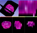
To enable Slicer 4 to load, view and process histopathology data.
Approach, Plan
Find, analyze and adapt the routines responsible for 3 channel image handling to enable processing of scanned histology slides with possible extensions towards registration of histo/path, digital pathology imaging and analysis.
Specifically we would like to be able:
- loading / saving of Slicer scenes that contain RGB volumes
- masking and segmentation of RGB volumes in Editor
Progress
Possible solutions have been identified (Downsampling, NRRD file format save) and applied. Loose ends have been identified.
Bug report submitted: http://www.na-mic.org/Bug/view.php?id=2225
Delivery Mechanism
This work will be delivered to the NA-MIC Kit as a (please select the appropriate options by noting YES against them below)
- Slicer Module
- Built-in YES
- Extension -- commandline NO
- Extension -- loadable PROBABLY
- Other (Please specify)
References
- Trivedi, H., B. Turkbey, et al. (2012). "Use of patient-‐specific MRI-‐based prostate mold for validation of multiparametric MRI in localization of prostate cancer." Urology 79(1): 233-‐239.
- "Elastic registration of multimodal prostate MRI and histology via multiattribute combined mutual information." Med Phys 38(4): 2005-‐2018.
- "Semi-‐automatic deformable registration of prostate MR images to pathological slices." J Magn Reson Imaging 32(5): 1149-‐1157.

