Difference between revisions of "NA-MIC Childrens Collaboration"
| (8 intermediate revisions by 2 users not shown) | |||
| Line 1: | Line 1: | ||
| − | Back to [[NA-MIC_External_Collaborations]] | + | __NOTOC__ |
| + | Back to [[NA-MIC_External_Collaborations|NA-MIC External Collaborations]] | ||
{| | {| | ||
| Line 7: | Line 8: | ||
|} | |} | ||
| − | + | ==Introduction== | |
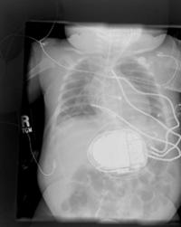
| − | Placement of Implantable Cardiac Defibrillators(ICDs)is a unique and challenging problem for children due to the variety of shapes and sizes, ranging from neonate to adolescent. As a result, a variety of novel implant techniques have been employed. Although these have generally been successful inasmuch as they result in a clinically acceptable defibrillation threshold, nothing is known about the mechanisms by which this threshold is attained, the optimal geometries for defibrillation, and whether unsafe electric field strengths are a result of novel implant approaches. Finite element modeling has been shown in adult torso models to correlate well with clinical results. Our goal is to model defibrillation in child torso models to gain insight into this important problem. We are also interested in developing new orientations in adults with the goal of lowering DFTs and providing options in patients with contraindications to standard techniques. | + | Placement of Implantable Cardiac Defibrillators(ICDs) is a unique and challenging problem for children due to the variety of shapes and sizes, ranging from neonate to adolescent. As a result, a variety of novel implant techniques have been employed. Although these have generally been successful inasmuch as they result in a clinically acceptable defibrillation threshold, nothing is known about the mechanisms by which this threshold is attained, the optimal geometries for defibrillation, and whether unsafe electric field strengths are a result of novel implant approaches. Finite element modeling has been shown in adult torso models to correlate well with clinical results. Our goal is to model defibrillation in child torso models to gain insight into this important problem. We are also interested in developing new orientations in adults with the goal of lowering DFTs and providing options in patients with contraindications to standard techniques. |
| − | == | + | ==Key Personnel== |
| + | *Stanford: Matthew Jolley, Anne Dubin, Paul Wang | ||
| + | *Children's Hospital Boston: John Triedman, Frank Cecchin | ||
| + | *Scientific Computing Institute, Utah: Jeroen Stinstra, Rob Macleod | ||
| + | *NorthEastern University: Dana Brooks | ||
| + | *NA-MIC: Steve Pieper, Kilian Pohl | ||
| − | + | = Goals of Project = | |
| − | + | ==Segmentation== | |
| − | + | Segmentation is currently done using tools within the open source tools 3D Slicer and Seg3D. We are actively working with members of SCI and SPL on developing automated segmentation techniques for torso and cardiac modeling. In the future we would like to integrate DT-MRI fiber structure into our cardiac models. | |
| − | == | + | ==Modeling== |
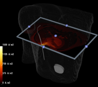
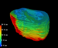
| − | + | We have worked with members of the SCI Institute in Utah to develop a modeling framework within their SCIRun package which allows rapid placement of realistic electrodes within finite element models of adult and child torsos. All tools to do so are available open source to download from the SCI website. We continue to improve this platform, integrating new technology and insight as we progress. | |
| + | |||
| + | ==Understanding Defibrillation== | ||
| + | Ultimately this project is part of a larger goal of understanding defibrillation as a therapy. Members of the group are also working with members of John Hopkins to further this goal. | ||
| − | + | = Building and Running SCIRun on SPL Machines = | |
| − | |||
| − | |||
| − | |||
| − | |||
[[SCRun on SPL Machines]] | [[SCRun on SPL Machines]] | ||
| − | == | + | ==Publications== |
| − | + | *Jolley M, Stinstra J, Weinstein D, et al. Open-Source Environment for Interactive Finite Element Modeling of Optimal ICD Electrode Placement Functional Imaging and Modeling of the Heart, Lecture Notes in Computer Science. Berlin/Heidelberg: Springer, 2007:373-382. | |
| − | + | *Jolley M, Stinstra J, Weinsten D, et al. Finite Element Modeling of Novel Defibrillation Approaches in Children and Adult Heart Rhythm Society. Denver, Colorado, 2007. | |
| − | + | *Jolley M., Triedman J., Westin C-F., Weinstein D.M., MacLeod R., Brooks D.H. [http://www.na-mic.org/publications/item/view/1240 Image Based Modeling of Defibrillation in Children.] Conf Proc IEEE Eng Med Biol Soc. 2006 Sep;1:2564-7. PMID: 17946966. | |
| − | + | * Stinstra J.G., Jolley M., Callahan M., Weinstein D., Cole M., Brooks D.H., Triedman J.K., Macleod R.S. [http://www.na-mic.org/publications/item/view/1241 Evaluation of Different Meshing Algorithms in the Computation of Defibrillation Thresholds in Children.] Conf Proc IEEE Eng Med Biol Soc. 2007 Aug;2007:1422-5. PMID: 18002232. | |
| − | + | *Jolley M., Stinstra J.G., Pieper S., Macleod R.S., Brooks D.H., Cecchin F., Triedman J.K. [http://www.na-mic.org/publications/item/view/1277 A Computer Modeling Tool for Comparing Novel ICD Electrode Orientations in Children and Adults.] Heart Rhythm. 2008 Apr;5(4):565-72. PMID: 18362024. PMCID: PMC2745086. | |
| − | |||
Latest revision as of 17:18, 14 December 2016
Home < NA-MIC Childrens CollaborationBack to NA-MIC External Collaborations
 
|
 
|
Introduction
Placement of Implantable Cardiac Defibrillators(ICDs) is a unique and challenging problem for children due to the variety of shapes and sizes, ranging from neonate to adolescent. As a result, a variety of novel implant techniques have been employed. Although these have generally been successful inasmuch as they result in a clinically acceptable defibrillation threshold, nothing is known about the mechanisms by which this threshold is attained, the optimal geometries for defibrillation, and whether unsafe electric field strengths are a result of novel implant approaches. Finite element modeling has been shown in adult torso models to correlate well with clinical results. Our goal is to model defibrillation in child torso models to gain insight into this important problem. We are also interested in developing new orientations in adults with the goal of lowering DFTs and providing options in patients with contraindications to standard techniques.
Key Personnel
- Stanford: Matthew Jolley, Anne Dubin, Paul Wang
- Children's Hospital Boston: John Triedman, Frank Cecchin
- Scientific Computing Institute, Utah: Jeroen Stinstra, Rob Macleod
- NorthEastern University: Dana Brooks
- NA-MIC: Steve Pieper, Kilian Pohl
Goals of Project
Segmentation
Segmentation is currently done using tools within the open source tools 3D Slicer and Seg3D. We are actively working with members of SCI and SPL on developing automated segmentation techniques for torso and cardiac modeling. In the future we would like to integrate DT-MRI fiber structure into our cardiac models.
Modeling
We have worked with members of the SCI Institute in Utah to develop a modeling framework within their SCIRun package which allows rapid placement of realistic electrodes within finite element models of adult and child torsos. All tools to do so are available open source to download from the SCI website. We continue to improve this platform, integrating new technology and insight as we progress.
Understanding Defibrillation
Ultimately this project is part of a larger goal of understanding defibrillation as a therapy. Members of the group are also working with members of John Hopkins to further this goal.
Building and Running SCIRun on SPL Machines
Publications
- Jolley M, Stinstra J, Weinstein D, et al. Open-Source Environment for Interactive Finite Element Modeling of Optimal ICD Electrode Placement Functional Imaging and Modeling of the Heart, Lecture Notes in Computer Science. Berlin/Heidelberg: Springer, 2007:373-382.
- Jolley M, Stinstra J, Weinsten D, et al. Finite Element Modeling of Novel Defibrillation Approaches in Children and Adult Heart Rhythm Society. Denver, Colorado, 2007.
- Jolley M., Triedman J., Westin C-F., Weinstein D.M., MacLeod R., Brooks D.H. Image Based Modeling of Defibrillation in Children. Conf Proc IEEE Eng Med Biol Soc. 2006 Sep;1:2564-7. PMID: 17946966.
- Stinstra J.G., Jolley M., Callahan M., Weinstein D., Cole M., Brooks D.H., Triedman J.K., Macleod R.S. Evaluation of Different Meshing Algorithms in the Computation of Defibrillation Thresholds in Children. Conf Proc IEEE Eng Med Biol Soc. 2007 Aug;2007:1422-5. PMID: 18002232.
- Jolley M., Stinstra J.G., Pieper S., Macleod R.S., Brooks D.H., Cecchin F., Triedman J.K. A Computer Modeling Tool for Comparing Novel ICD Electrode Orientations in Children and Adults. Heart Rhythm. 2008 Apr;5(4):565-72. PMID: 18362024. PMCID: PMC2745086.