Difference between revisions of "Project Week 25/CNN for multi-plane prostate segmentation"
From NAMIC Wiki
| Line 40: | Line 40: | ||
==Illustrations== | ==Illustrations== | ||
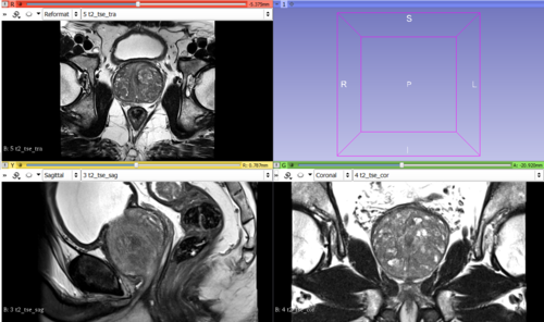
| − | [[File:MRIImage.png|thumb|MRIs of the prostate (axial, sagittal and coronal volume)]] | + | {| |
| − | [[File:Segmentation2.png|thumb| | + | |[[File:MRIImage.png|thumb|500px|MRIs of the prostate (axial, sagittal and coronal volume)]] |
| − | + | |- | |
| + | |[[File:Segmentation2.png|thumb|500px|Example segmentation (apex, mid-gland and base slices).]] | ||
| + | |} | ||
==Background and References== | ==Background and References== | ||
<!-- Use this space for information that may help people better understand your project, like links to papers, source code, or data --> | <!-- Use this space for information that may help people better understand your project, like links to papers, source code, or data --> | ||
Latest revision as of 11:42, 30 June 2017
Home < Project Week 25 < CNN for multi-plane prostate segmentation
Back to Projects List
Key Investigators
- Anneke Meyer (University of Magdeburg, Germany)
- Alireza Mehrtash (Brigham and Women's Hospital, Harvard Medical School, USA)
- Christian Hansen (University of Magdeburg, Germany)
- Andrey Fedorov (Brigham and Women's Hospital, Harvard Medical School, USA)
- Jennifer Nitsch (University of Bremen, Germany)
Project Description
| Objective | Approach and Plan | Progress and Next Steps |
|---|---|---|
|
For prostate lesion assessment, MRI volumes in sagittal, coronal and axial orientation are acquired. The goal is to segment the prostate in these volumes and fuse the segmentations in order to obtain better accuracy, especially in the apex and base regions (compared to segmentations obtained only on axial volumes). |
|
|

