Difference between revisions of "Projects:DiffeoMixedEffects"
Jfishbaugh (talk | contribs) (Created page with 'Back to Utah 2 Algorithms __NOTOC__ = Ongoing Work = == Diffeomorphic Shape Trajectories for Improved Longitudinal Segmentation and Statistics == Longitudi…') |
Jfishbaugh (talk | contribs) |
||
| Line 8: | Line 8: | ||
Longitudinal image data has several sources of variability. First, there is inherent biological variability, both within a subject changing over time and also between subjects in a population. The goal of longitudinal analysis is to quantify this variability and make inferences about changes over time in a population. However, longitudinal imaging data also include unwanted sources of variability, such as noise in image acquisition, segmentation and registration errors, and human expert judgment, among others. These extraneous errors tend to dampen statistical power, especially when trying to distinguish between trajectories of two different populations, e.g., healthy and diseased. | Longitudinal image data has several sources of variability. First, there is inherent biological variability, both within a subject changing over time and also between subjects in a population. The goal of longitudinal analysis is to quantify this variability and make inferences about changes over time in a population. However, longitudinal imaging data also include unwanted sources of variability, such as noise in image acquisition, segmentation and registration errors, and human expert judgment, among others. These extraneous errors tend to dampen statistical power, especially when trying to distinguish between trajectories of two different populations, e.g., healthy and diseased. | ||
| − | [[File:Caudate_volume_plot.png|800px|center|thumb|'''Fig. | + | [[File:Caudate_volume_plot.png|800px|center|thumb|'''Fig. 3.''' Left: For one subject, volume of observed caudates (open circles) and temporally consistent continuous volume extracted from diffeomorphic shape model (solid line). The difference in caudate volume extracted from scans obtained on the same day highlights the need for consistent segmentation. Right: Observed volume and volume extracted from diffeomorphic shape models for all 65 subjects. While the volume of the discrete shape observations show considerable variation, volume extracted from personalized models are continuous and temporally consistent.]] |
| − | [[File:Observed_vs_consistent_striatal_volume.png|800px|center|thumb|'''Fig. | + | [[File:Observed_vs_consistent_striatal_volume.png|800px|center|thumb|'''Fig. 2.''' Longitudinal mixed-effects analysis of striatal volumes obtained from observed shapes (Left) and temporally consistent shapes (Right). Volume data are shown as filled black circles with corresponding individual trends. Note the improvement of the model fit in the consistent striatal volume over the observed striatal volume, which results in lower standard error of estimated fixed-effects parameters.]] |
| + | |||
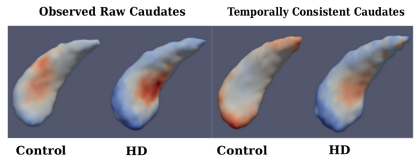
| + | [[File:Compare_caudates.png|600px|center|thumb|'''Fig. 3.''' Left: Fixed-effects parameters for observed caudate shapes (Far Left-Control, Mid Left- HD), Right: Fixed-effects parameters for temporally consistent caudate shapes (Mid Right-Control, Far Right-HD); Fixed effects slope: Blue-Red indicates Local Contraction - Expansion.]] | ||
= Literature = | = Literature = | ||
Revision as of 05:44, 14 October 2014
Home < Projects:DiffeoMixedEffectsBack to Utah 2 Algorithms
Ongoing Work
Diffeomorphic Shape Trajectories for Improved Longitudinal Segmentation and Statistics
Longitudinal image data has several sources of variability. First, there is inherent biological variability, both within a subject changing over time and also between subjects in a population. The goal of longitudinal analysis is to quantify this variability and make inferences about changes over time in a population. However, longitudinal imaging data also include unwanted sources of variability, such as noise in image acquisition, segmentation and registration errors, and human expert judgment, among others. These extraneous errors tend to dampen statistical power, especially when trying to distinguish between trajectories of two different populations, e.g., healthy and diseased.


Literature
[1] Muralidharan, P., Fishbaugh, J., Johnson, H., Durrleman, S., Paulsen, J., Gerig, G., Fletcher, P.T. Diffeomorphic shape trajectories for improved longitudinal segmentation and statistics. Proc. of Medical Image Computing and Computer Assisted Intervention (MICCAI '14). (2014).
Key Investigators
- Utah: Prasanna Muralidharan, James Fishbaugh, P.T. Fletcher, Guido Gerig
- INRIA/ICM, Pitie Salpetriere Hospital: Stanley Durrleman
- Department of Psychiatry, Carver College of Medicine, University of Iowa: Jane Paulsen, Hans Johnson
