Difference between revisions of "Projects:RegistrationLibrary:RegLib C29"
From NAMIC Wiki
| Line 26: | Line 26: | ||
===Objective / Background === | ===Objective / Background === | ||
This is a classic case of a multi-sequence MRI exam we wish to spatially align to the anatomical reference scan (T1-SPGR). The scan of interest is the DTI image to be aligned for surgical planning/reference. | This is a classic case of a multi-sequence MRI exam we wish to spatially align to the anatomical reference scan (T1-SPGR). The scan of interest is the DTI image to be aligned for surgical planning/reference. | ||
| + | |||
| + | === Download === | ||
| + | *DATA | ||
| + | **[[Media:RegLib_C29_DATA.zip|'''Registration Library Case 29 (MRI) <small> (Data & Solution Xforms, incl. DTI but not DWI or resampled DTI, zip file 73 MB) </small>]] | ||
| + | **[[Media:RegLib_C29_DATA2.zip|'''Registration Library Case 29 (MRI) <small> (DWI and resampled DTI images, zip file 198 MB) </small>]] | ||
| + | *PRESETS: | ||
| + | *TUTORIALS: | ||
| + | **[[Media:RegLib_C29_Screencast.mov| Screencast movie (Quicktime, 73 MB)]] | ||
| + | **[[Media:RegLib_C29_DTI.ppt| Powerpoint Tutorial (.ppt PowerPoint file, 1.5 MB)]] | ||
| + | **[[Media:RegLib_C29_DTI.pdf| Powerpoint Tutorial as PDF (PDF file 2.1 MB)]] | ||
=== Keywords === | === Keywords === | ||
| Line 114: | Line 124: | ||
[[Image:RegLib_C29_AGif_full.gif|500px|Registered DTI superimposed on SPGR and T2]] registered (cycles show T1 and T2 and color DTI overlay)<br> | [[Image:RegLib_C29_AGif_full.gif|500px|Registered DTI superimposed on SPGR and T2]] registered (cycles show T1 and T2 and color DTI overlay)<br> | ||
<br> | <br> | ||
| − | |||
| − | |||
| − | |||
| − | |||
| − | |||
| − | |||
| − | |||
| − | |||
| − | |||
| − | |||
Revision as of 22:12, 14 September 2010
Home < Projects:RegistrationLibrary:RegLib C29Back to ARRA main page
Back to Registration main page
Back to Registration Use-case Inventory
Contents
v3.6.1  Slicer Registration Library Case #29: Intra-subject Brain DTI
Input
|
|
|
|

|
|
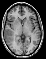
| fixed image/target T1 SPGR |
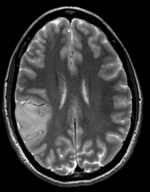
moving image 1 T2 |
moving image 2a DTI baseline |
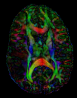
moving image 2b DTI tensor |
Modules
- Slicer 3.6.1 recommended modules: BrainsFit
Objective / Background
This is a classic case of a multi-sequence MRI exam we wish to spatially align to the anatomical reference scan (T1-SPGR). The scan of interest is the DTI image to be aligned for surgical planning/reference.
Download
- DATA
- PRESETS:
- TUTORIALS:
Keywords
MRI, brain, head, intra-subject, DTI, T1, T2, non-rigid, tumor, surgical planning
Input Data
- reference/fixed : T1 SPGR , 0.5x0.5x1 mm voxel size, 512 x 512 x 176
- moving 1: T2 0.5x0.5x1.5 mm voxel size, 512 x 512 x 92
- moving 2a: DTI baseline: 1 x 1 x 3 mm, 256 x 256 x 41
- moving 2b: 1 x 1 x 3 mm, 256 x 256 x 41 x 9 (tensor), original: DWI 256 x 256 x 41 x 32 directions
Registration Challenges
- The DTI sequence (EPI) contains string distortions we seek to correct via non-rigid alignment
- the DTI baseline is similar in contrast to a T2, albeit at much lower resolution
- we have different amounts of voxel-anisotropy
- a direct registration of the DTI_baseline to the SPGR will fail, hence a 2-step approach is required
Key Strategies
- Slicer 3.6.1 recommended modules: BrainsFit
- to align the DTI with the T1 we need 2 registration steps: 1.align the T2 with the T1 and 2. align the DTI with the T2. The DTI baseline scan has contrast most similar to the T2 and hence is best registered against the T2. However the structural reference scan with highest resolution and tissue contrast is the T1.
- we therefore use the following approach: 1) we first co-register the T2 with the SPGR T1. 2) we register the DTI baseline to the registered/resampled T2; 3) resample the DTI volume with the new transform
- the DTI-T2 registration includes non-rigid deformation to correct for the strong distortions from the EPI acquisition. Because of the nonrigid component a mask of the brain parenchyma helps greatly in obtaining a meaningful transform.
- The DTI estimation provides an automated mask for the DTI_baseline scan, but we have no mask for the T2. We can either obtain one through separate segmentation or by sending the DTI_mask through an additional registration step. In this example we show the latter.
- thus the full pipeline is this:
- Affine align T2-T1, incl. resampled T2 volume = T2r
- Affine+BSpline align of DTI_baseline to T2r (unmasked)
- Resample DTI_mask with above BSpline -> mask for the T2r
- repeat Affine+BSpline align of DTI_base to T2r, WITH masks
- resample DTI with result Affine+BSpline transform
Procedures
- Phase I: LOAD DATA
- download example dataset
- load into 3DSlicer 3.6.1 (Load Scene)
- To convert the DWI into a DTI: use the Converters / DICOM to NRRD Converter module
- Phase II: Register T2 to SPGR
- open Registration : BrainsFit module
- Registration Phases:
- select/check Include Rigid registration phase
- select/check Include Affine registration phase
- select a new transform Output Transform
- Registration Parameters: increase Number Of Samples to 200,000
- Leave all other settings at default
- click apply; runtime ca. 1-2 min.
- Resample T2 into T1 space
- Open Resample Scalar/Vector/DWI Volume module (Filtering menu)
- Input Volume: T2, Reference Volume: T1
- Output Volume: create new volume, rename to "T2_Xf1"
- Interpolation Type: select ws (windowed sinc)
- Click Apply.
- Upon completion, go to Volumes module to adjust window & level
- Active Volume: select T2_Xf1
- Open Display tab and adjust window & level, e.g. 1300/700
- Phase III:REGISTER DTI TO T2_Xf1
- open Registration : BrainsFit module
- Registration Phases:
- set T2_Xf1 as fixed and DTI_baseline as moving image
- select/check Include Rigid registration phase
- select/check Include Affine registration phase
- select/check Include BSpline registration phase
- select Initialize with CenterofHeadAlign
- select Include Rigid registration phase
- select "Include Affine registration phase"
- Output Settings:
- select a new transform "Slicer BSpline Transform", rename to "Xf2_DTI-T1
- select a new volume "Output Image Volume
- Registration Parameters: increase Number Of Samples to 200,000
- Leave all other settings at default
- click apply; runtime ca. 3 min.
- Phase IV: Resample DTI
- Load the combined transform (Add Data)
- Open the Resample DTI Volume module (found under: All Modules)
- Input Volume: select DTI
- Output Volume: select New DTI Volume, rename to DTI_Xf2
- Reference Volume: select T1
- Transform Parameters: select transform "Xf2_DTI-T1
- check box: output-to-input
- Leave all other settings at defaults
- Click Apply; runtime ca. 3-4 min.
- Go to the Volumes module, select the newly produced DTI_Xf2 volume
- under the Display tab, select Color Orientation from the Scalar Mode menu
- set T1 as background and new DTI_Xf2 volume as foreground
- Set fade slider to see DTI overlay onto the SPGR image
for more details see the tutorial(s) under Downloads
Registration Results
Registered DTI superimposed on SPGR and T2 registered (cycles show T1 and T2 and color DTI overlay)