Difference between revisions of "Projects:TubularSurfaceSegmentationPopStudy"
| Line 1: | Line 1: | ||
| − | Back to [[Algorithm: | + | Back to [[Algorithm:BU|Boston University Algorithms]] |
__NOTOC__ | __NOTOC__ | ||
= Group study using the Tubular Surface model = | = Group study using the Tubular Surface model = | ||
Revision as of 16:11, 12 January 2012
Home < Projects:TubularSurfaceSegmentationPopStudyBack to Boston University Algorithms
Group study using the Tubular Surface model
Description
We have proposed a new framework for performing group studies on DW-MRI data using the Tubular Surface Model to study white-matter properties. We show that the model facilitates population studies by the natural registration that occurs by the sampling of WM properties along the fiber bundles center-lines. Further, by allowing us to characterize the discrimination ability of local regions of the fiber bundles, the framework allows us to identify the regions that are "affected" by the disorder under study. In our experiments, we have applied the framework to study the Cingulum Bundle towards discriminating Schizophrenia.
== Some Results The results below show the visualization of t-statistics with respect to the extracted features, with discrimination ability increasing from green to red. This demonstrates the ability of the framework to visualize the role of different regions of the fiber bundle in Schizophrenia.
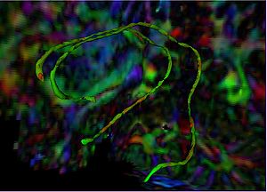 Visualization of T-statistics on Cingulum Bundle surface (View 1)
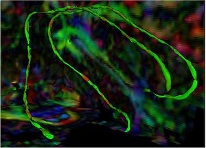 Visualization of T-statistics on Cingulum Bundle surface (View 2)


Key Investigators
Georgia Tech: Allen Tannenbaum, Vandana Mohan
BWH: Marek Kubicki
Publications
In Print
In Press
V. Mohan, G. Sundaramoorthi, M. Kubicki and A. Tannenbaum. Population Analysis of neural fiber bundles towards schizophrenia detection and characterization, using the Tubular Surface model. Neuroimage (in submission)