Difference between revisions of "Rhesus WMGM Atlas"
From NAMIC Wiki
| Line 8: | Line 8: | ||
* Using a rhesus tissue atlas provided by the UNC Neuro Image Analysis Laboratory and the UWisc Harlow Primate Laboratory we have segmented the data into WM/GM, and CSF capartments using the Lobulated EM Segmentation method. These segmentations have been averaged to create a study-specific tissue atlas. | * Using a rhesus tissue atlas provided by the UNC Neuro Image Analysis Laboratory and the UWisc Harlow Primate Laboratory we have segmented the data into WM/GM, and CSF capartments using the Lobulated EM Segmentation method. These segmentations have been averaged to create a study-specific tissue atlas. | ||
| − | <gallery caption="Rhesus Probabilistic Atlas" widths="350px" heights="180px" perrow=" | + | <gallery caption="Rhesus Probabilistic Atlas" widths="350px" heights="180px" perrow="1"> |
Image:T1template_Cr.jpg|Population Specific T1 template image | Image:T1template_Cr.jpg|Population Specific T1 template image | ||
Image:T2template_cr.jpg|Population Specific T2 template image | Image:T2template_cr.jpg|Population Specific T2 template image | ||
Revision as of 20:10, 6 February 2008
Home < Rhesus WMGM AtlasObjective:
- Develop an adult WM/GM/CSF atlas from normal subject images.
Progress:
- N=17 T1,T2 subject images acquired
- Using a rhesus tissue atlas provided by the UNC Neuro Image Analysis Laboratory and the UWisc Harlow Primate Laboratory we have segmented the data into WM/GM, and CSF capartments using the Lobulated EM Segmentation method. These segmentations have been averaged to create a study-specific tissue atlas.
- Rhesus Probabilistic Atlas
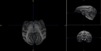
Population Specific T1 template image
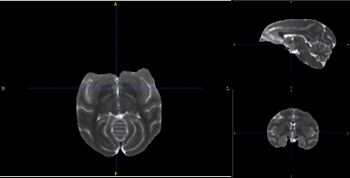
Population Specific T2 template image
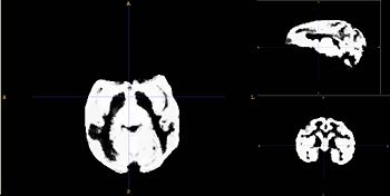
Probabilistic map of GM for Rhesus population
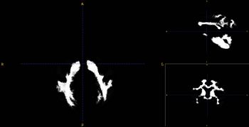
Probabilistic map of WM for Rhesus population


Key Investigators:
- Wake Forest: Bob Kraft, Jim Daunais
- Virginia Tech: Chris Wyatt