Difference between revisions of "Slicer3:VisualBlog"
| Line 2: | Line 2: | ||
<gallery caption="2008" widths="200px" perrow="4"> | <gallery caption="2008" widths="200px" perrow="4"> | ||
| + | Image:Cardioseg+volume.png|'''Cardiac segmentation and CT Volume Rendering, February 2008'''<br> Using data and segmentations from the [[NA-MIC_Childrens_Collaboration | collaboration with Boston Children's Hospital Pediatric Cardiology.]]. | ||
Image:Slicer_IGTL_PartialImage.png|'''Partial image update using OpenIGTLink, February 2008'''<br> [[OpenIGTLink]] allows an external imaging scanner (Ultrasound/CT/MR) to update part of a volume, which has already been loaded on the Slicer. This is helpful if the imaging plane sweeps the subject, and images are transfered to the Slicer on-the-fly. | Image:Slicer_IGTL_PartialImage.png|'''Partial image update using OpenIGTLink, February 2008'''<br> [[OpenIGTLink]] allows an external imaging scanner (Ultrasound/CT/MR) to update part of a volume, which has already been loaded on the Slicer. This is helpful if the imaging plane sweeps the subject, and images are transfered to the Slicer on-the-fly. | ||
Image:Slicer_CudaHead.jpg|'''The First MRML node rendering using CUDA February 2008'''<br> [[Slicer3:Volume_Rendering_With_Cuda|Cuda Volume Rendering]] provides a Volume Rendering Method on the new and advanced NVidia Hardware. | Image:Slicer_CudaHead.jpg|'''The First MRML node rendering using CUDA February 2008'''<br> [[Slicer3:Volume_Rendering_With_Cuda|Cuda Volume Rendering]] provides a Volume Rendering Method on the new and advanced NVidia Hardware. | ||
Revision as of 13:56, 12 February 2008
Home < Slicer3:VisualBlog
- 2008
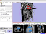
Cardiac segmentation and CT Volume Rendering, February 2008
Using data and segmentations from the collaboration with Boston Children's Hospital Pediatric Cardiology..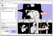
Partial image update using OpenIGTLink, February 2008
OpenIGTLink allows an external imaging scanner (Ultrasound/CT/MR) to update part of a volume, which has already been loaded on the Slicer. This is helpful if the imaging plane sweeps the subject, and images are transfered to the Slicer on-the-fly.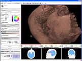
The First MRML node rendering using CUDA February 2008
Cuda Volume Rendering provides a Volume Rendering Method on the new and advanced NVidia Hardware.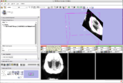
The First Implementation of OpenIGTLink, January 2008
OpenIGTLink protocol provides plug-and-play connectivity to tracking devices, imagers (MR, ultrasound ...) and other medical devices. A real-time image transfer to the Slicer in 10 fps is demonstrated in the screenshot.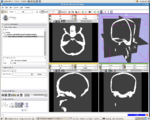
Image from Haiying Liu, Patrick Cheng, Noby Hata, Junichi Tokuda, Luis Ibanez, and Steve Pieper, January 2008.
In the NAMIC All Hands Meeting 2008, the bi-directional socket communication has been established between the Tracker Daemon in Slicer 3 and IGSTK server which acquires tracking data from device and sends it to Tracker Daemon. The speed of communication is controlled by Slicer.


- 2007
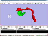
Image from Jaesung Hong December 2007
Visualization of cochlea(green) and facial nerve(red) for ENT navigation. We are moving on from Slicer2 to Slicer3 at Kyushu University Hospital in Japan.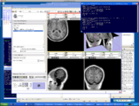
Image from Steve Pieper, Luis Ibanez, Haiying Liu December 2007
Result of Slicer for IGT workshop showing Slicer3 transform node being updated by a IGSTK tracker process.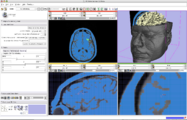
Image from Steve Pieper, Andy Freudling December 2007
More volume rendering examples. New Threshold tool makes it easy to visualize surfaces.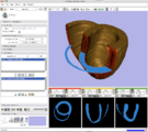
Image from Steve Pieper, December 2007
Display of segmented heart data results using volume rendering inside Slicer3. See JHU Computational Anatomy Portal] for more information.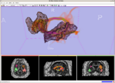
Image from Tri Ngo and Steve Pieper, November 2007
Display of Stochastic Tractography results using volume rendering inside Slicer3. See here for more information.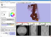
Image from Andy Freudling on October 2007
First results of volume rendering inside Slicer3. See here for more information.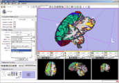
Image from wjp on Wednesday, October 24, 2007
QueryAtlas visualizing combined morphology and functional analyses generated by running FreeSurfer and a FIPS pipeline on a BIRN phaseII SIRP dataset. Interactive annotations (on mouse-over) are being translated thru Slicer's controlled vocabulary.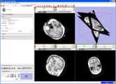
Image from Jim on Saturday, September 15, 2007
Slice viewers can be used to specify oblique reformats using the 'Reformat' orientation (instead of axial, coronal, sagittal) and CTRL-Right-Button-Move
(subject to change).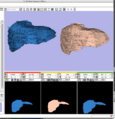
Image from Dirk, Matthew, Jim, and Steve on Monday, August 13, 2007
A new label map smoothing tool has been added to help with our collaboration with Children's Hospital Boston, SCI at University of Utah and Northeastern University. The unfiltered labelmap is shown in blue, and the filtered results are shown in peach.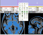
Image from Steve and Wendy on Monday, August 6, 2007
New Editor functionality, with EditBox which is invoked using the F1 key (will soon be moved to the space bar).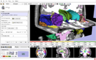
Image from Brad and Kilian on Wednesday, June, 21, 2007
Example of EMSegmenter in Slicer3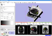
Image from pieper on Friday, June, 9, 2007
Slicer3 Module for Stochastic Tractography from MIT (Ngo, Golland) and BWH (Westin, Kubicki).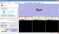
Image from ipek on Wednesday, June, 6, 2007
UNC Logo in Slicer3 for KWMeshVisu (Ipek Oguz, Martin Styner).
Image from wjp on Wednesday, May, 30, 2007
Design mockups for Slicer3's Cine Display interface (William Leue, Wendy Plesniak).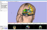
Image from pieper on Tuesday, May, 30, 2007
The SPL-PNL brain atlas loaded in Slicer3. Demonstrates model hierarchy and clipping.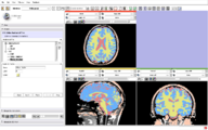
Image from davisb on Monday, April, 27, 2007
EMSegment screenshot---segmentation results and work-flow GUI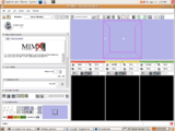
Image from magnotta on Monday, April, 16, 2007
Mimx Logo in Slicer3 for VoxelMeshingModule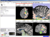
Image from wjp on Thursday, March 22, 2007 at 10:00AM
New GUI elements: Shows a magnified view in the GUI panel of an area around the mouse in any Slice Window.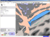
Image from wjp on Thursday, March 22, 2007 at 10:00AM
New GUI elements: Shows birds-eye-view of the scene relative to the outline of the 3D Viewer's window in the 'Manipulate 3D View' GUI panel.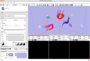
Image from pieper on Tuesday, January 16, 2007 at 7:58PM
From Lauren, shows fiber tracts loaded from a file and with display properties controlled by the GUI.


- 2006
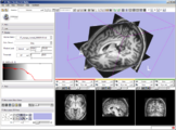
Image from wjp on Friday, December 29, 2006 at 3:23PM
Module choose and navigation functionality integrated into the application toolbar to create more space for module GUIs on the side panel. Slice Controller widgets also updated to have more functionality available, a button to link and unlink their control, and an improved visual design.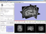
Image from wjp on Monday, November 28, 2006 at 6:40M
New 3D Slicer logo integrated with slicer GUI.
Image from pieper on Thursday, October 26, 2006 at 1:54M
FreeSurfer structural data (cortical and subcortical segmentation) with fBIRN functional overlay, shown as part of Slicer3-based BIRN Query Atlas project.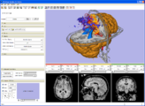
Image from pieper on Thursday, October 05, 2006 at 1:08PM
Clipping with the slice plane is now supported, along with thresholded image display.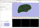
Image from naucoin on Friday, September 08, 2006 at 12:14pm
The FreeSurfer colour lookup table has been added to the Slicer3 FreeSurfer Library. A curvature overlay file was loaded and displayed on bert/surf/lh.pial as loaded in via the Models module.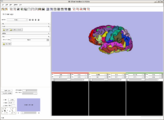
Image from naucoin on Friday, September 08, 2006 at 11:24am
The FreeSurfer scalar reader has been integrated as a Slicer 3 Library, and surfaces can be read via the Models module. Shot shows standard bert/surf/lh.pial plus a test script to load the annotation labels file.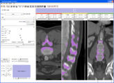
Image from pieper on Tuesday, August 08, 2006 at 4:26PM
A number of things have come together to allow the new prototype editor module. More ...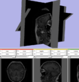
Image from pieper on Friday, July 28, 2006 at 9:04AM
Test of making a non-orthogonal slice plane. This was created by manually setting the SliceToRAS matrix in the vtkMRMLSliceNode. GUI is not yet hooked up for oblique slices.
Image from pieper on Thursday, July 27, 2006 at 5:21PM
Shows that a single pixel voxel in black is exactly bounded by a unit cube in RAS space. Also the blinking eye buttons for slice visiblility are hooked up and there is now a red-yellow-green color coding for the slice windows (carried over from slicer2).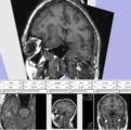
Image from pieper on Thursday, July 13, 2006 at 4:49PM
Test of interactive slice textured plane control. Not yet eneabled through GUI, but driven by a test script that you can source into the console. More ...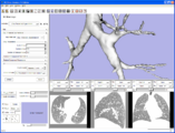
Image from pieper on Tuesday, July 11, 2006 at 3:39PM
Example model read from vtk file and loaded in slicer3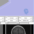
Image from pieper on Monday, July 03, 2006 at 3:35PM
Test of the new logo widget for adding module-specific watermark logos directly in the 3d view. Uses a vtkLogoWidget from VTK cvs head. Kitware is working on cmake support to notify developers when a VTK cvs update is needed. Once that is done, the logo code will be added to slicer3 svn.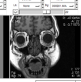
Image from pieper on Monday, June 26, 2006 at 10:38AM
New annotations and key bindings. Middle mouse to pan, right mouse to zoom.
Image by pieper on Wednesday, June 21, 2006 at 1:01PM
results of the command line median filter module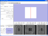
Image by millerjv on Tuesday, June 20, 2006 at 4:37PM
{First command line module to export data, execute, and import data back into Slicer}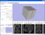
Image by millerjv on Friday, June 16, 2006 at 11:15AM
{Command line module with an enumerated parameter as a radio button}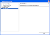
Image by millerjv on Wednesday, June 14, 2006 at 10:20AM
{Slicer applications settings panel showing the module search path}
Image from pieper on Tuesday, June 13, 2006 at 8:23PM
Another command line module example from Jim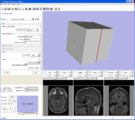
Image from pieper on Tuesday, June 13, 2006 at 8:20PM
Example from Jim Miller showing the automatic GUI generation from XML collected from execution module commands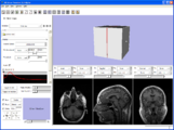
Image from pieper on Tuesday, June 13, 2006 at 8:18PM
Example screen shot showing that there is a basic interface in place.


About the VisualBlog
The VisualBlog is meant to be an easy place to upload screenshot so that both developers and outside observes can track the progress of the project.
Many thanks to developers and users for contributing their images here.