File list
From NAMIC Wiki
This special page shows all uploaded files.
| Date | Name | Thumbnail | Size | Description | Versions |
|---|---|---|---|---|---|
| 00:51, 9 November 2011 | Septal Reg Movie CARMA.mov (file) | 1.14 MB | A short movie showing a linear blend from the registered pre-ablation DEMRI scan to the post-ablation scan. The main region of misregistration in this image pair is the septal wall of the LA. | 1 | |
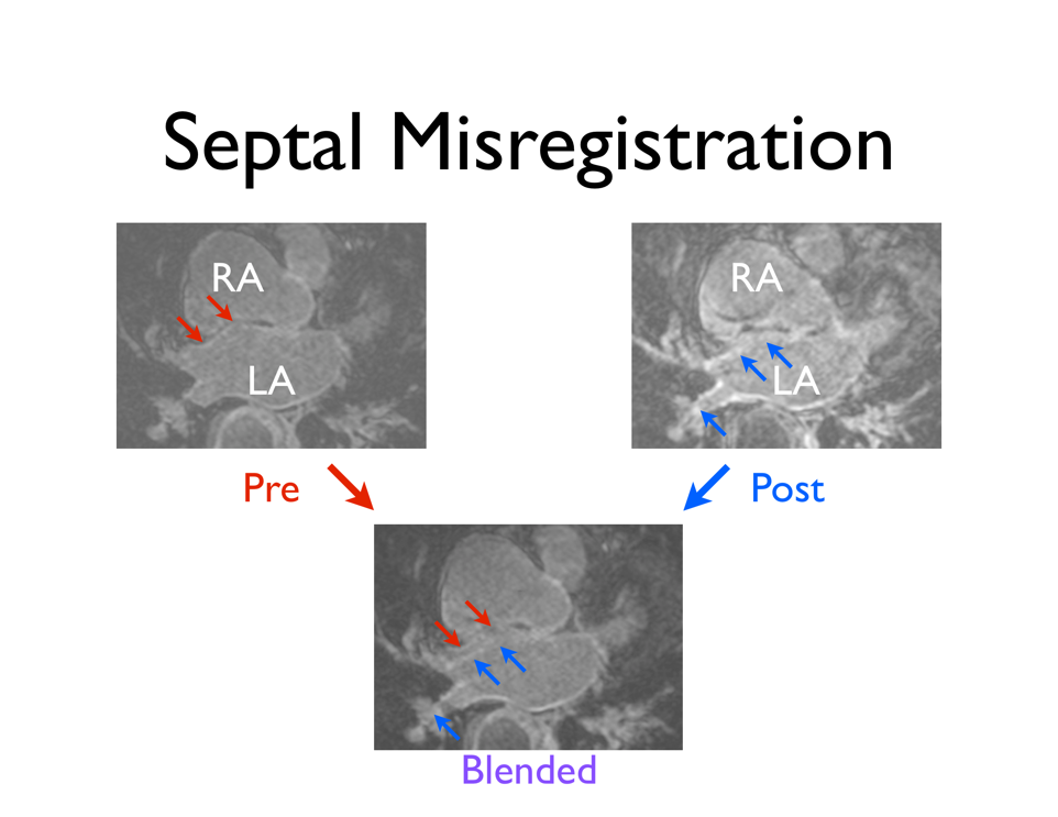
| 00:23, 9 November 2011 | Septal MisReg CARMA.png (file) |  |
224 KB | An example of the pre- and post-ablation images from the same patient that were registered with the BrainsFit module. The registration was good except along the septal surface (arrows). | 1 |
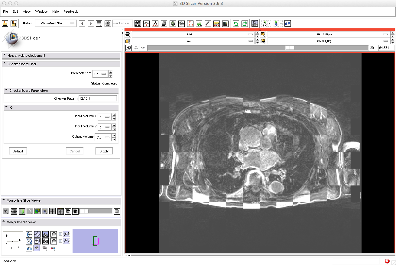
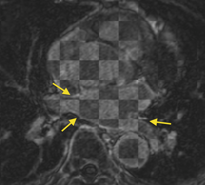
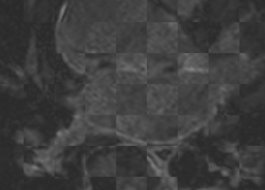
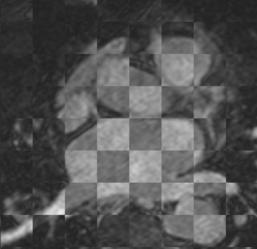
| 21:13, 9 November 2011 | Septal Checker CARMA.png (file) |  |
403 KB | Checkerboard filtered image of an image where the septum of the LA was misregistered between the pre- and 3 month post-ablation images. | 1 |
| 00:49, 9 November 2011 | PV Reg Movie CARMA.mov (file) | 1.74 MB | 2 | ||
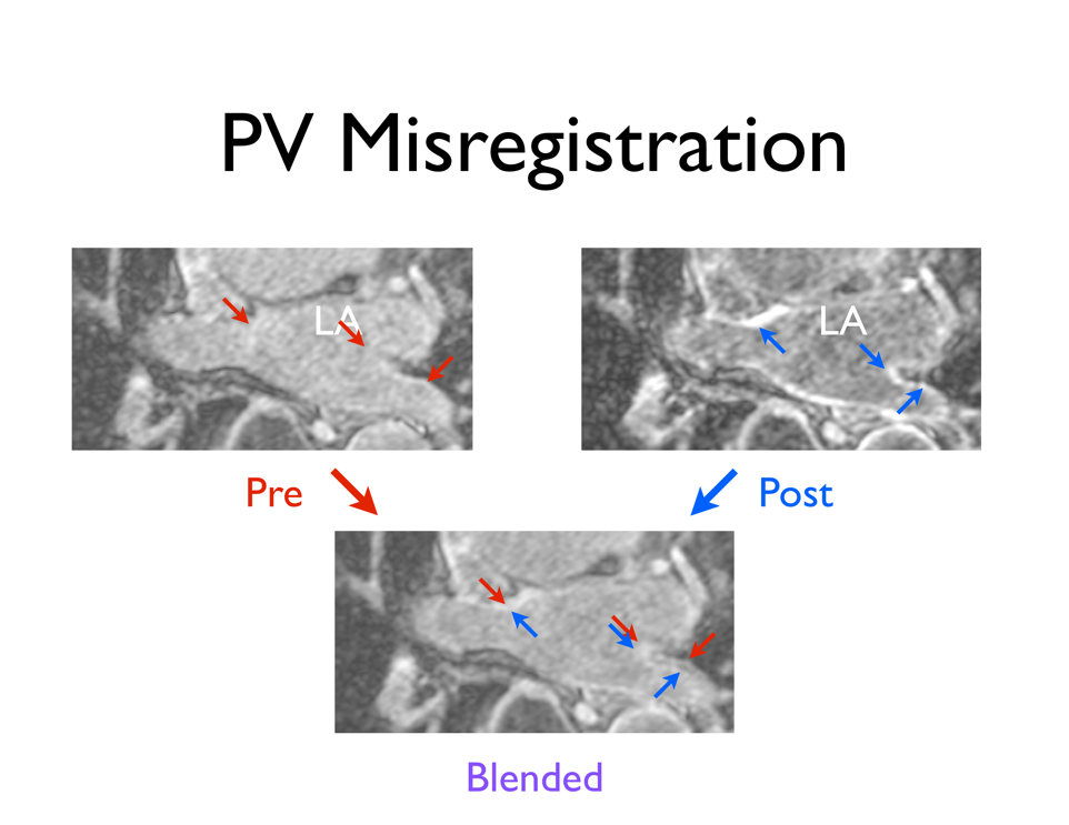
| 00:27, 9 November 2011 | PV MisReg CARMA.png (file) |  |
229 KB | Affine and rigid registration (BrainsFit module) of the same patient's pre- and post-ablation scans improved the alignment of the PVs, but sufficient overlap of the corresponding PV structures was not achieved. | 1 |
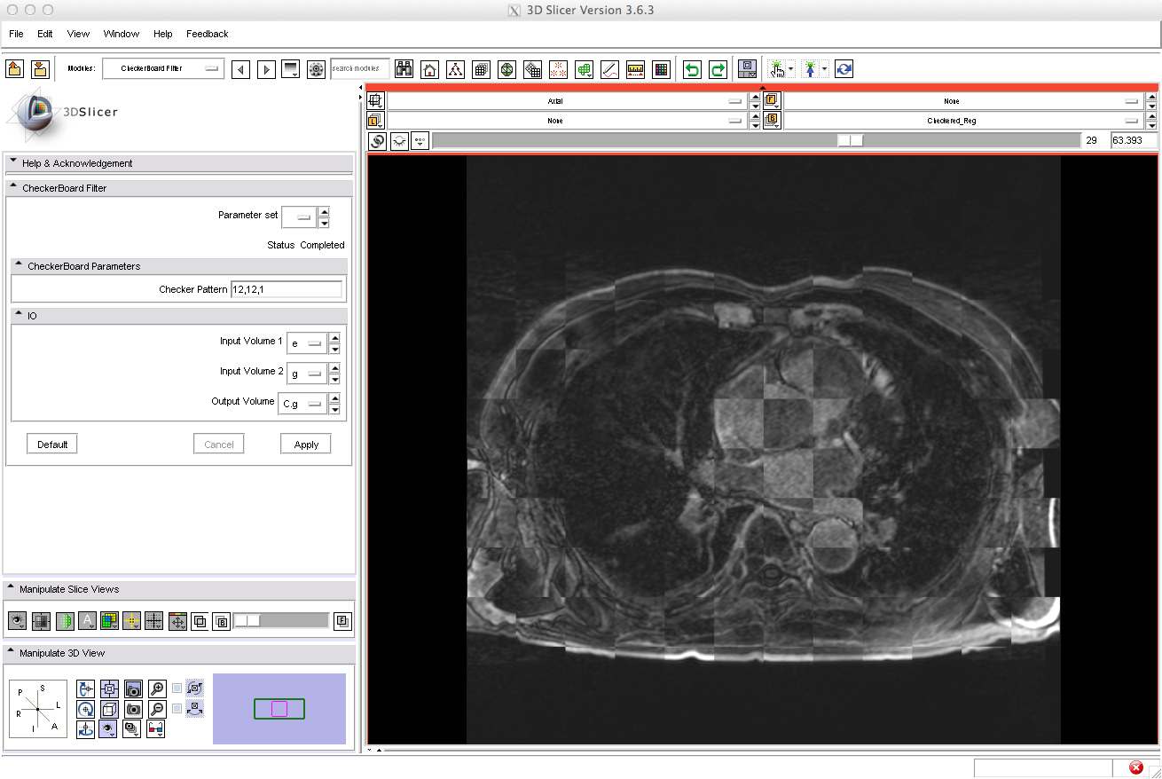
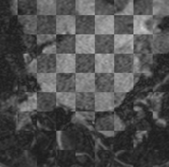
| 21:12, 9 November 2011 | PV Checker CARMA.png (file) |  |
402 KB | A checkerboard filter derived image of registered pre- and post-ablation images showing poor alignment of the PVs. | 1 |
| 04:03, 30 August 2011 | NAMIC Flow Chart.png (file) |  |
683 KB | Proposed CARMA Registration Pipeline. This diagrams the proposed pipeline for registering the pre- and post-ablation images. The current state (8.29.11) of the Slicer code is denoted by the red, dashed bounding box. | 1 |
| 03:54, 30 August 2011 | NAMIC Flow Chart.pdf (file) | 2.18 MB | The proposed workflow. The raw image stacks are processed through a series of transformations, cropping, and MRI bias correction. The pipeline yields cropped, registered pre- and post-ablation volumes. The transformation matrix of the registration step | 2 | |
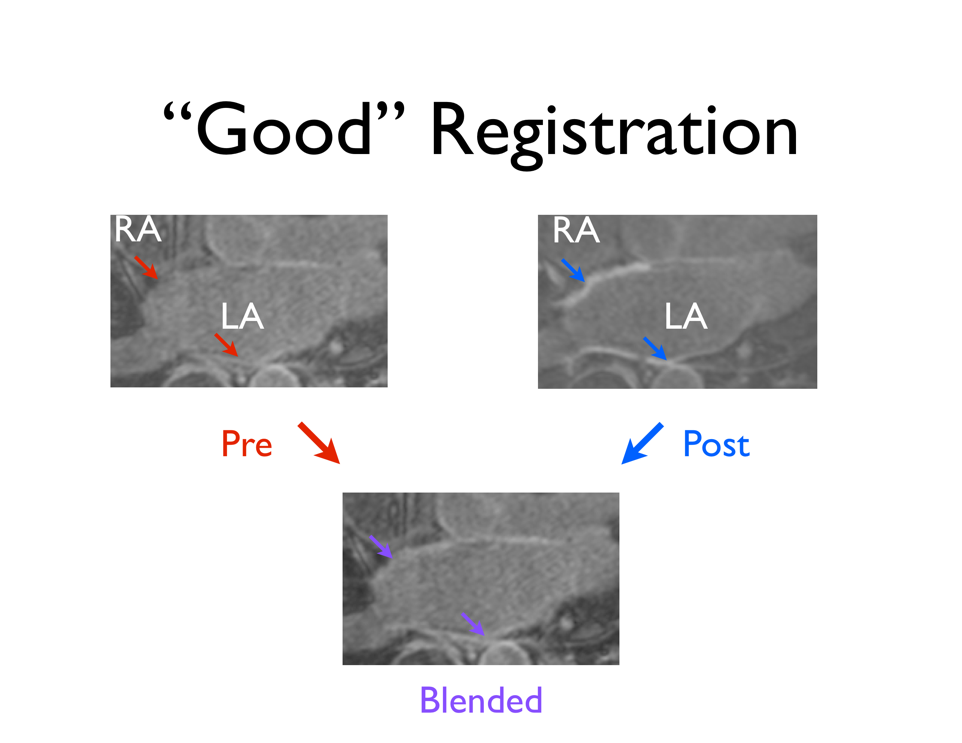
| 00:19, 9 November 2011 | Good Registration CARMA.png (file) |  |
165 KB | An example of generally good registration of both the LA wall and PVs on a patient's pre- and 3 month post-ablation DEMRI scans. | 1 |
| 00:11, 9 November 2011 | Good Registration CARMA.pdf (file) | 79 KB | An example of generally good registration of both the LA wall and PVs on a patient's pre- and 3 month post-ablation DEMRI scans. | 1 | |
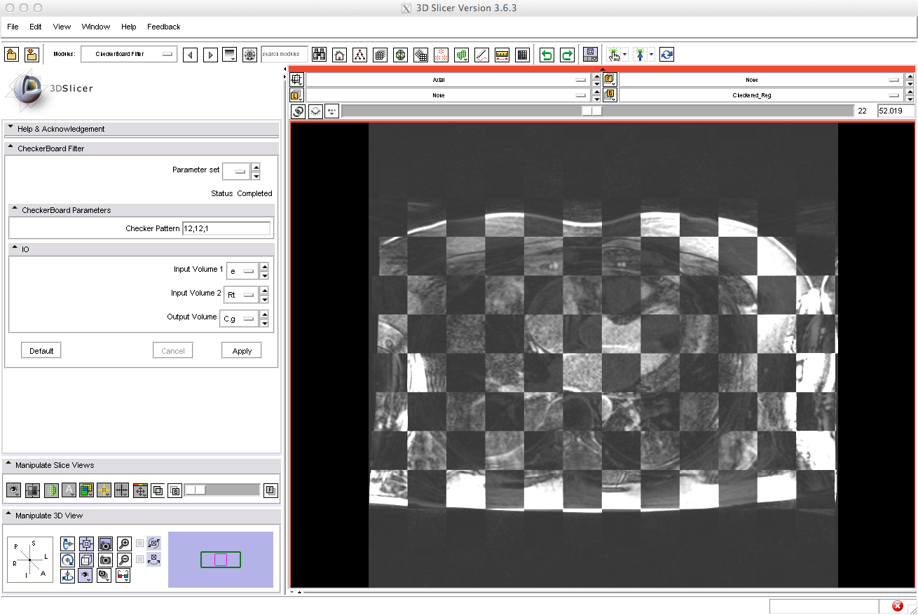
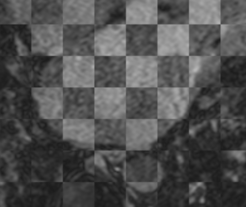
| 21:09, 9 November 2011 | Good Checker CARMA.png (file) |  |
394 KB | A combination of the registered whole-body pre- and post-ablation DEMRI images, where the lighter checkers represent the pre-ablation image. Good registration of the LA structures was seen. | 1 |
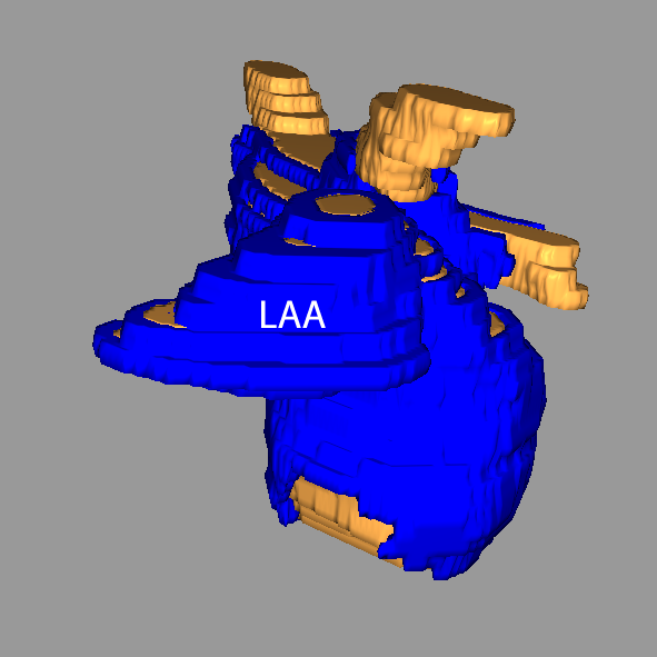
| 00:51, 9 January 2012 | Carma vert LAA.png (file) |  |
148 KB | 2 | |
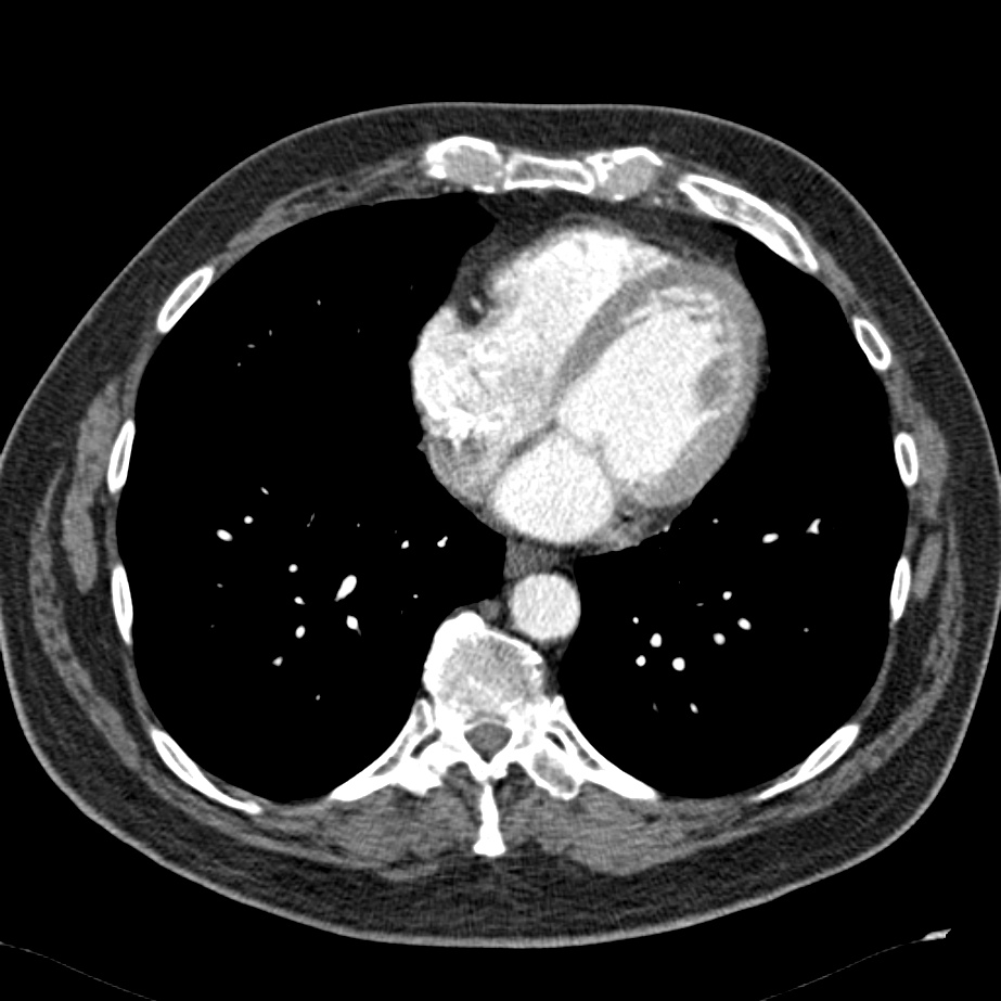
| 18:20, 10 May 2012 | Carma reg CT.jpg (file) |  |
170 KB | Example CARMA CT image (likely DynaCT). | 1 |
| 00:52, 9 January 2012 | Carma norm LAA.png (file) |  |
98 KB | Reverted to version as of 00:51, 9 January 2012 | 4 |
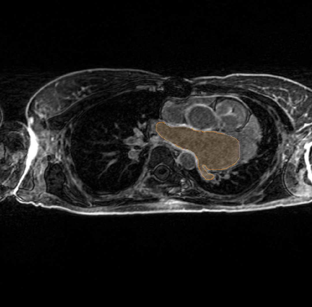
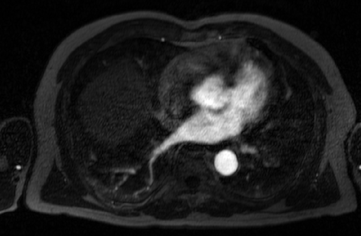
| 00:30, 9 January 2012 | Carma no reflow.png (file) |  |
327 KB | LGE-MRI scans acquired immediately post-ablation pose extra challenges for registration; often times, edema and other acute physiological changes effect the washout kinetics of the contrast agent and the morphology of the LA. Here the MR contrast agent h | 1 |
| 18:20, 10 May 2012 | Carma no reflow.jpg (file) |  |
170 KB | Example CARMA CT image (likely DynaCT). | 1 |
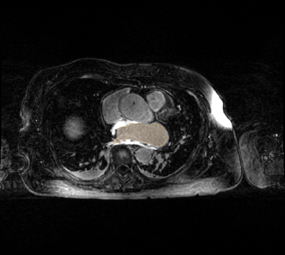
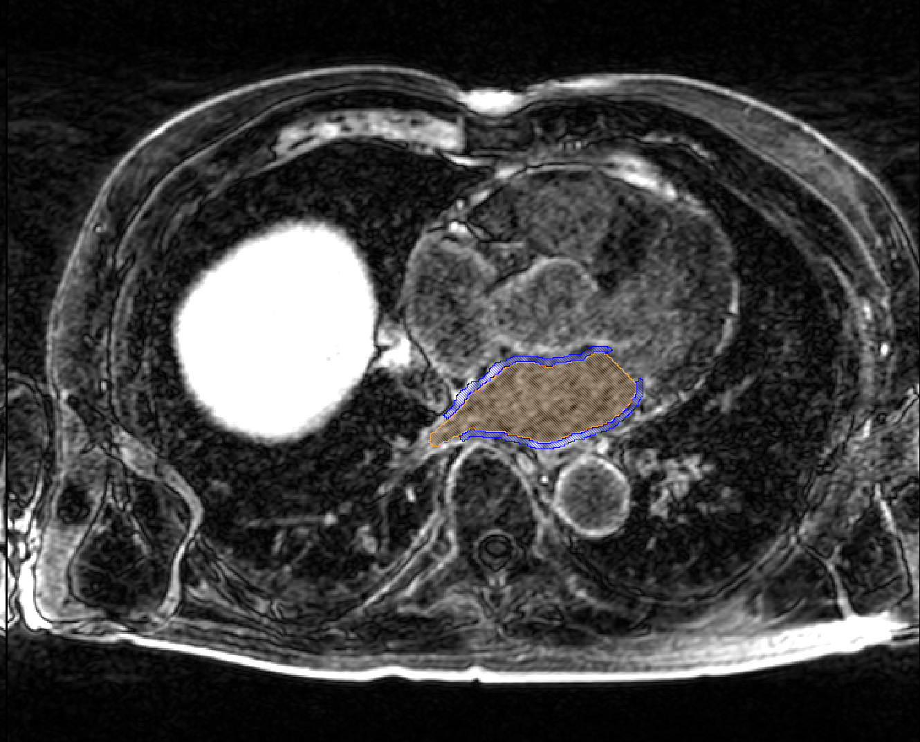
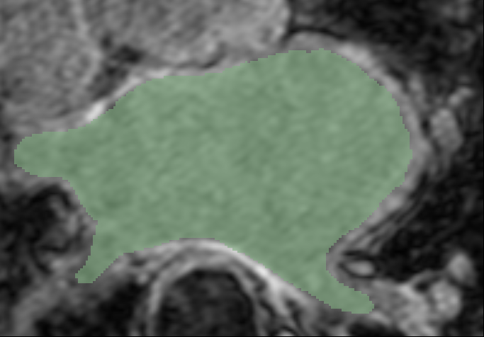
| 00:24, 9 January 2012 | Carma hypertrophy.png (file) |  |
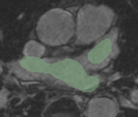
303 KB | An example of a hypertrophied LA, the extent of which is denoted by the orange label mask. | 1 |
| 17:47, 9 January 2012 | Carma fid misreg.png (file) |  |
243 KB | 2 | |
| 22:43, 6 January 2012 | Carma ex pre seg.png (file) |  |
388 KB | Carma Center example pre-ablation LGE-MRI image, with manually segmented LA wall and blood pool overlaid | 1 |
| 22:44, 6 January 2012 | Carma ex pre.png (file) |  |
267 KB | Carma Center example pre-ablation LGE-MRI image | 1 |
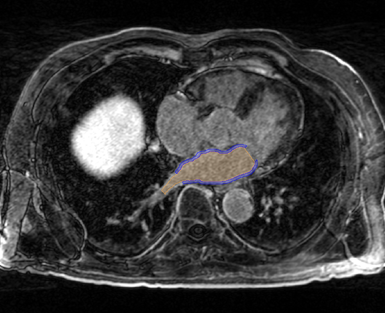
| 22:42, 6 January 2012 | Carma ex post seg.png (file) |  |
393 KB | Carma Center example post-ablation LGE-MRI image, with manual segmentations of the LA wall and blood pool overlaid | 1 |
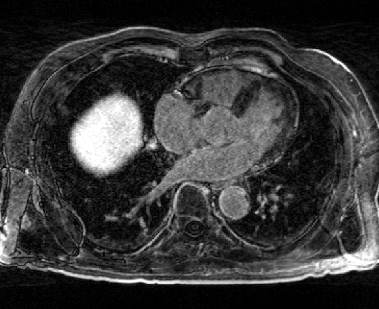
| 22:43, 6 January 2012 | Carma ex post.png (file) |  |
276 KB | Carma Center example post-ablation LGE-MRI image | 1 |
| 22:41, 6 January 2012 | Carma ex mra.png (file) |  |
179 KB | Carma Center example MRA image (acquired prior to LGE-MRI image) | 1 |
| 23:49, 6 January 2012 | Carma RPVs small.png (file) |  |
130 KB | Variations in the size of the PVs also occurs; here the inferior PV is dramatically smaller than normal and the superior larger than is typical. | 1 |
| 17:42, 9 January 2012 | Carma PV misreg.png (file) |  |
268 KB | An example of the misregistration that is seen when trying to register the pulmonary veins of pre- and post-ablation LGE-MRI scans from the same patient (scans acquired roughly 3 months apart). | 1 |
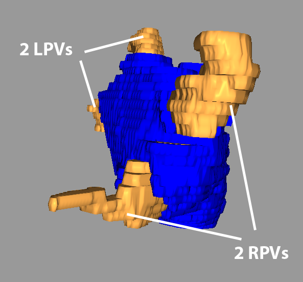
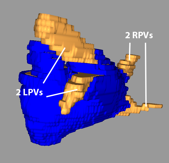
| 00:00, 7 January 2012 | Carma Norm RPVs.png (file) |  |
115 KB | A typical arrangement of the PVs: 2 distinct LPVs and 2 RPVs. | 1 |
| 00:00, 7 January 2012 | Carma Norm LPVs.png (file) |  |
138 KB | A typical arrangement of the PVs: 2 distinct LPVs and 2 RPVs. | 1 |
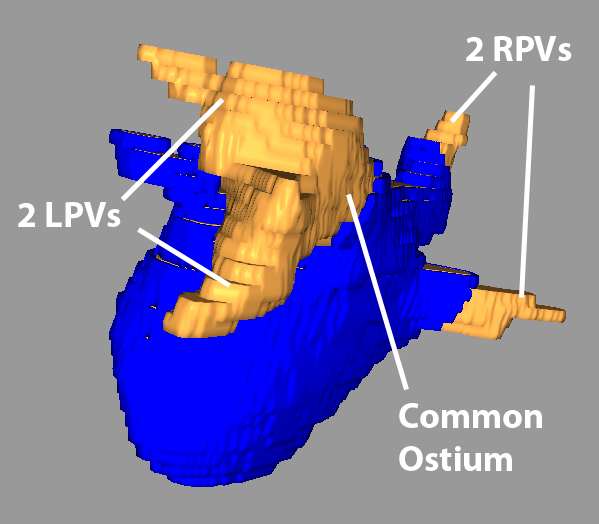
| 23:47, 6 January 2012 | Carma Com LPV.png (file) |  |
122 KB | A LA with the typical number of PVs (2 left, 2 right); however, the 2 left PVs share a common trunk. | 1 |
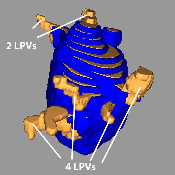
| 23:46, 6 January 2012 | Carma 4rpvs.png (file) |  |
165 KB | A LA with the 2 left PVs (typical) and an abnormal 4 right PVs. | 1 |
| 23:22, 8 January 2012 | Carma 4RPV.png (file) |  |
193 KB | A LA with the 2 left PVs (typical) and an abnormal 4 right PVs. | 1 |
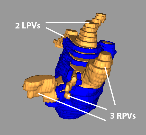
| 23:03, 8 January 2012 | Carma 3RPVs.png (file) |  |
148 KB | Reverted to version as of 00:13, 7 January 2012 | 4 |
| 23:25, 8 January 2012 | Carma 3RPV.png (file) |  |
148 KB | A LA with the 2 left PVs (typical) and an abnormal 3 right PVs. | 1 |
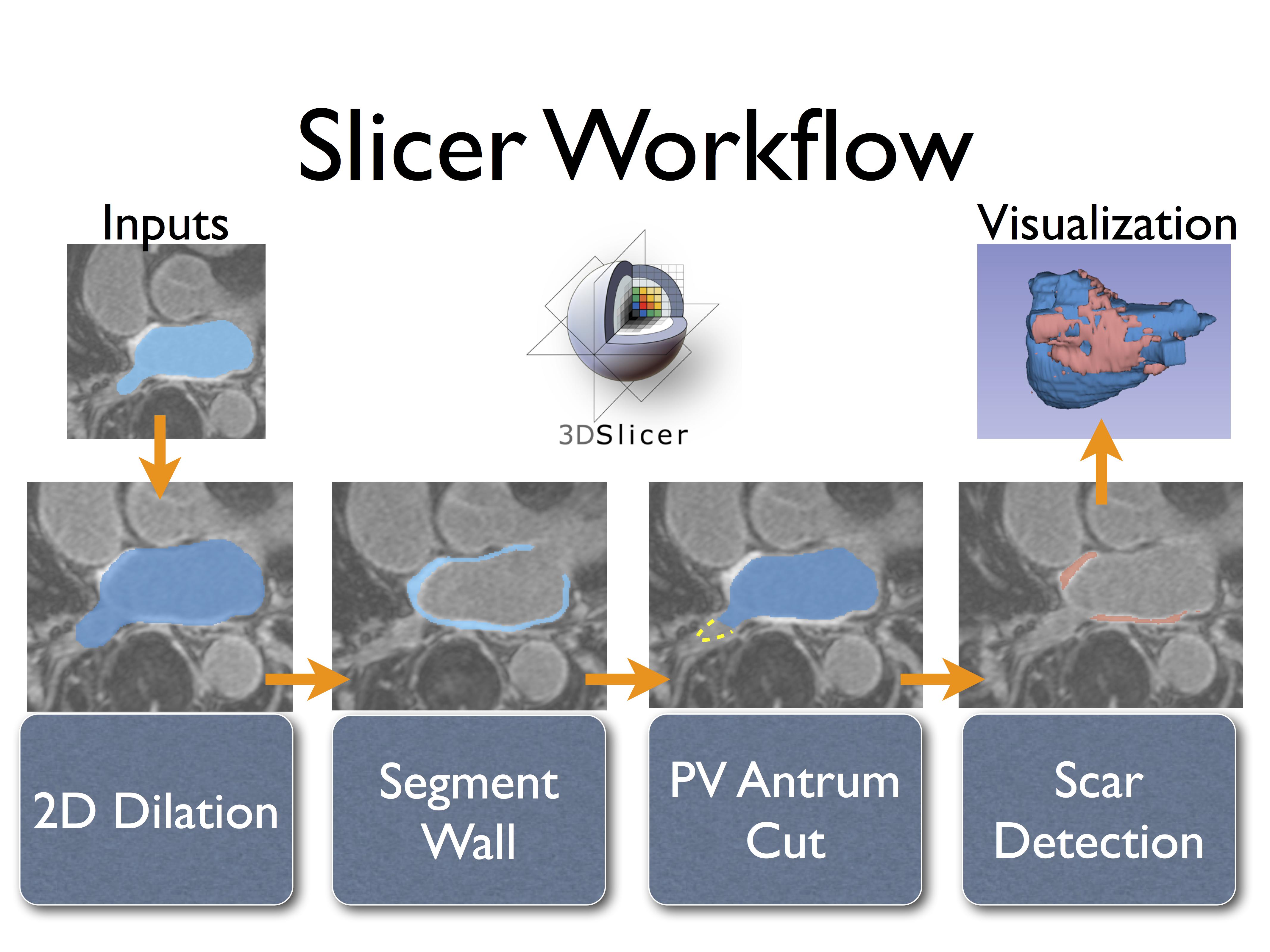
| 07:24, 10 January 2013 | CarmaWorkflowV2.pdf (file) | 4.71 MB | Overview of the CARMA fibrosis/scar segmentation workflow in Slicer. | 1 | |
| 07:20, 10 January 2013 | CarmaWorkflowSlide.png (file) | Error creating thumbnail: File with dimensions greater than 12.5 MP |
3.94 MB | Overview of the CARMA fibrosis segmentation workflow in Slicer. | 3 |
| 07:15, 10 January 2013 | CarmaWorkflowSlide.pdf (file) | 792 KB | Overview of the CARMA scar detection algorithm in Slicer. | 1 | |
| 07:29, 10 January 2013 | CarmaWorkflowSlide.jpeg (file) |  |
546 KB | Overview of the CARMA fibrosis/scar segmentation workflow in Slicer. | 1 |
| 06:22, 3 April 2012 | CARMA VecReg Seg3.png (file) |  |
113 KB | 1 | |
| 06:23, 3 April 2012 | CARMA VecReg Seg3-2.png (file) |  |
98 KB | 1 | |
| 06:38, 3 April 2012 | CARMA VecReg Seg2.png (file) |  |
55 KB | 2 | |
| 06:18, 3 April 2012 | CARMA VecReg Seg1.png (file) |  |
106 KB | 1 | |
| 06:18, 3 April 2012 | CARMA VecReg Seg1-2.png (file) |  |
77 KB | 1 | |
| 06:38, 3 April 2012 | CARMA VecReg Check3.png (file) |  |
63 KB | 2 | |
| 06:22, 3 April 2012 | CARMA VecReg Check2.png (file) |  |
139 KB | 1 | |
| 06:22, 3 April 2012 | CARMA VecReg Check2-2.png (file) |  |
95 KB | 1 | |
| 06:17, 3 April 2012 | CARMA VecReg Check1.png (file) |  |
116 KB | 1 | |
| 06:19, 3 April 2012 | CARMA VecReg Check1-2.png (file) |  |
125 KB | 1 | |
| 08:25, 29 November 2011 | CARMA UT Endo.png (file) |  |
223 KB | Segmentation of an atlas image by Utah. | 1 |
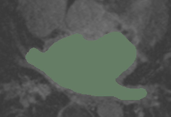
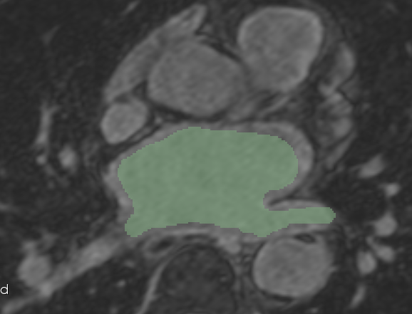
| 08:43, 29 November 2011 | CARMA N26 LGE.png (file) |  |
260 KB | Auto-segmentation mask and cropped LGE image example. | 1 |
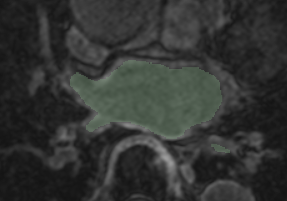
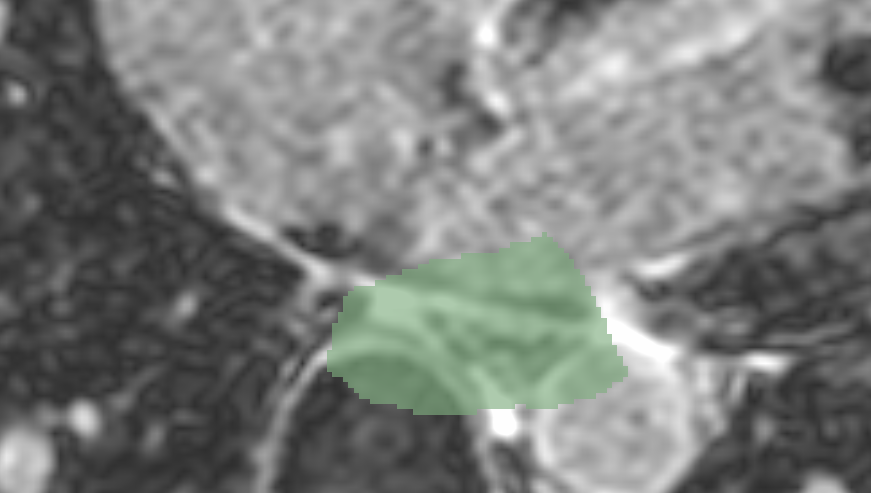
| 08:45, 29 November 2011 | CARMA N26 Endo.png (file) |  |
13 KB | Auto-segmentation mask (green) and the expert-defined manual segmentation (white) of the blood pool. | 1 |
| 08:43, 29 November 2011 | CARMA N26 2 LGE.png (file) |  |
295 KB | Auto-segmentation mask and cropped LGE image example. | 1 |