File list
From NAMIC Wiki
This special page shows all uploaded files.
| Date | Name | Thumbnail | Size | Description | Versions |
|---|---|---|---|---|---|
| 14:07, 24 June 2011 | Posterior base 4 class.png (file) |  |
2 KB | 1 | |
| 14:10, 24 June 2011 | Posterior follow class 4.png (file) |  |
3 KB | 1 | |
| 14:07, 24 June 2011 | Posterior base 5 class.png (file) |  |
4 KB | 1 | |
| 14:10, 24 June 2011 | Posterior follow class 3.png (file) |  |
7 KB | 1 | |
| 14:07, 24 June 2011 | Posterior base 3 class.png (file) |  |
8 KB | 1 | |
| 14:06, 24 June 2011 | Posterior base 1 class.png (file) |  |
8 KB | 1 | |
| 14:10, 24 June 2011 | Posterior follow class 1.png (file) |  |
9 KB | 1 | |
| 14:10, 24 June 2011 | Posterior follow class 2.png (file) |  |
12 KB | 1 | |
| 14:07, 24 June 2011 | Posterior base 2 class.png (file) |  |
12 KB | 1 | |
| 05:02, 30 September 2011 | Vector mag 2D.png (file) |  |
15 KB | 1 | |
| 05:57, 9 January 2012 | DefVis VecMag TBISeg.png (file) |  |
15 KB | 1 | |
| 14:00, 24 June 2011 | Ori follow Channel3 Slice 69.png (file) |  |
17 KB | 1 | |
| 05:03, 30 September 2011 | Jacobian det 2D.png (file) |  |
19 KB | 1 | |
| 05:54, 9 January 2012 | DefVis DetJacobian TBISeg.png (file) |  |
19 KB | 2 | |
| 13:50, 24 June 2011 | Ori base Channel3 Slice 69.png (file) |  |
20 KB | 1 | |
| 18:15, 7 January 2013 | DWI sample subject.png (file) |  |
20 KB | 1 | |
| 00:25, 6 January 2014 | Active 4D.png (file) |  |
25 KB | 1 | |
| 17:10, 20 June 2011 | Wm sup seg namic 11.png (file) |  |
26 KB | 1 | |
| 12:09, 13 January 2012 | New data p4 3.png (file) |  |
26 KB | 1 | |
| 13:54, 22 June 2012 | Vis of ventrical.jpg (file) |  |
27 KB | 1 | |
| 12:09, 13 January 2012 | New data p4 2.png (file) |  |
27 KB | 1 | |
| 13:50, 24 June 2011 | Ori base Channel2 Slice 69.png (file) |  |
28 KB | 1 | |
| 14:00, 24 June 2011 | Ori follow Channel2 Slice 69.png (file) |  |
28 KB | 1 | |
| 13:51, 24 June 2011 | Ori base Channel4 Slice 69.png (file) |  |
31 KB | 1 | |
| 14:00, 24 June 2011 | Ori follow Channel4 Slice 69.png (file) |  |
31 KB | 1 | |
| 13:50, 24 June 2011 | Ori base Channel1 Slice 69.png (file) |  |
34 KB | 1 | |
| 14:00, 24 June 2011 | Ori follow Channel1 Slice 69.png (file) |  |
35 KB | 1 | |
| 04:48, 30 September 2011 | Seg algorithm framework.png (file) | 36 KB | 2 | ||
| 12:08, 13 January 2012 | New data p4 1.png (file) |  |
37 KB | 1 | |
| 14:00, 21 June 2013 | UIPlanPic2.png (file) |  |
37 KB | 1 | |
| 06:44, 9 January 2012 | TBI Seg lable Slicer.png (file) |  |
39 KB | 1 | |
| 04:51, 30 September 2011 | Joint analysis flowchart.png (file) |  |
40 KB | 1 | |
| 18:15, 7 January 2013 | MRI sample subject.png (file) |  |
48 KB | 1 | |
| 13:20, 13 January 2012 | Semiauto seg 2D X view.jpg (file) |  |
65 KB | 1 | |
| 13:20, 13 January 2012 | Manual seg 2D X view.jpg (file) |  |
65 KB | 1 | |
| 06:01, 21 June 2013 | UIPlanPic.png (file) |  |
82 KB | 1 | |
| 06:47, 14 January 2011 | WM surfaces TBI P3 time2.png (file) |  |
108 KB | 1 | |
| 06:46, 14 January 2011 | WM surfaces TBI P3 time1.png (file) |  |
108 KB | 1 | |
| 06:45, 14 January 2011 | Top view patient3 time1.png (file) |  |
113 KB | 1 | |
| 11:06, 11 January 2013 | Diff reg TBI.png (file) |  |
114 KB | 1 | |
| 06:45, 14 January 2011 | Bottom view patient3 time1.png (file) |  |
115 KB | 1 | |
| 06:47, 14 January 2011 | Top view patient3 time2.png (file) |  |
117 KB | 1 | |
| 06:47, 14 January 2011 | Bottom view patient3 time2.png (file) |  |
117 KB | 1 | |
| 05:45, 16 June 2012 | Atlas contruction illu TBI.png (file) |  |
124 KB | 1 | |
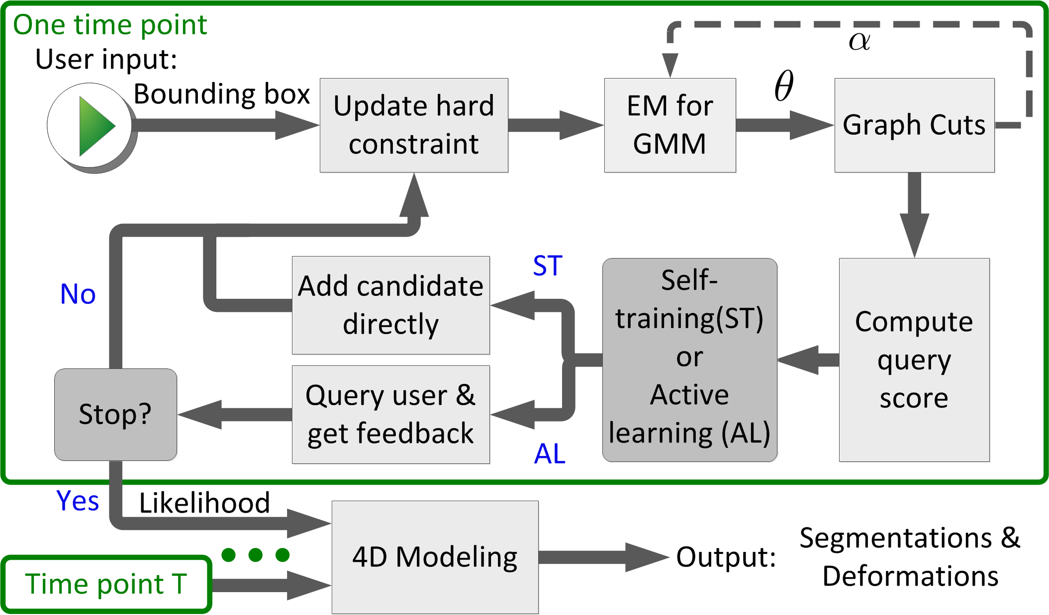
| 21:33, 11 October 2014 | Flowchart 4D active cut.png (file) |  |
141 KB | 1 | |
| 12:27, 13 January 2012 | Seg P4 followup result 3D topview sp.png (file) |  |
146 KB | 1 | |
| 12:20, 13 January 2012 | Seg P4 followup result 3D leftview sp.png (file) |  |
147 KB | 1 | |
| 03:46, 27 June 2014 | TBI active seg.jpg (file) |  |
155 KB | 1 | |
| 06:06, 12 September 2011 | One subject chronic seg.png (file) |  |
231 KB | 1 | |
| 01:11, 17 December 2010 | Coregistration mulmodalities u.png (file) |  |
251 KB | 1 |