File list
From NAMIC Wiki
This special page shows all uploaded files.
| Date | Name | Thumbnail | Size | Description | Versions |
|---|---|---|---|---|---|
| 17:41, 28 April 2008 | CA anAutomaton.png (file) |  |
10 KB | depict a cellular automaton | 1 |
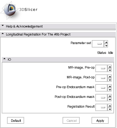
| 17:45, 12 January 2012 | AFibLongitudinalRegGUI.png (file) |  |
13 KB | 1 | |
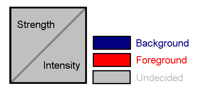
| 21:14, 1 December 2008 | Posterior.png (file) |  |
14 KB | distance modulated posterior map | 1 |
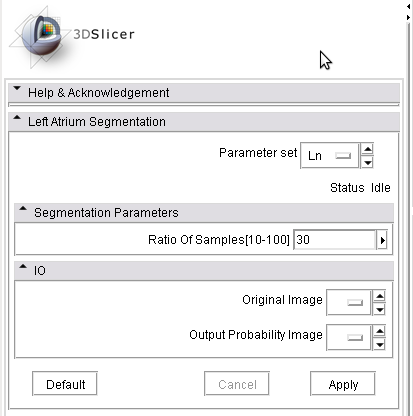
| 15:16, 14 January 2011 | LAsegmentationModuleUI.png (file) |  |
14 KB | 1 | |
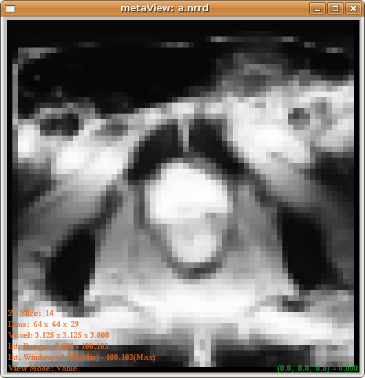
| 17:00, 1 March 2011 | RSS Grayscale-label.nrrd (file) | 16 KB | 1 | ||
| 16:53, 1 March 2011 | MORSS Grayscale-multilabel.nrrd (file) | 16 KB | 1 | ||
| 15:39, 1 March 2011 | RssPanel.png (file) |  |
17 KB | 1 | |
| 23:38, 1 December 2008 | PosteriorWithUniformPrior.png (file) |  |
23 KB | posterior with uniform prior | 1 |
| 14:13, 8 March 2011 | RSS sub2 cardix LVlabel.nrrd (file) | 24 KB | 1 | ||
| 14:56, 7 June 2013 | SegAidedRegSquareFocus128.png (file) |  |
25 KB | Icon for the SegmentationAidedRegistration extension | 1 |
| 14:20, 8 March 2011 | MORSS CARDIX-sub2-multilabel.nrrd (file) | 26 KB | 1 | ||
| 03:50, 25 June 2010 | Hippo480-s-crop.png (file) |  |
37 KB | 1 | |
| 03:49, 25 June 2010 | Hippo480-a-crop.png (file) |  |
41 KB | 1 | |
| 03:50, 25 June 2010 | Hippo480-c-crop.png (file) |  |
42 KB | 1 | |
| 21:52, 12 January 2012 | AFibEndoSegAtlasAndRSS sagittal.png (file) |  |
68 KB | 1 | |
| 21:52, 12 January 2012 | AFibEndoSegAtlasAndRSS coronal.png (file) |  |
69 KB | 1 | |
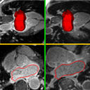
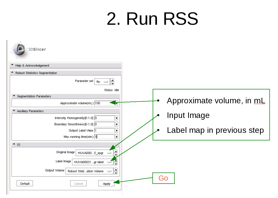
| 01:55, 8 April 2010 | RobustStatisticsSegmentation usage2.png (file) |  |
87 KB | 3 | |
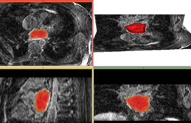
| 19:41, 12 June 2008 | Prostate20080318 seg.png (file) |  |
87 KB | The segmentation of prostate using Random Walk algorithm. | 2 |
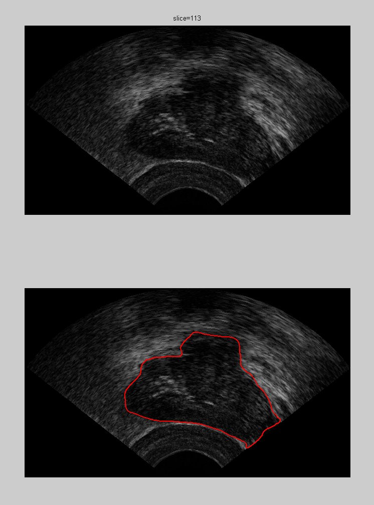
| 18:51, 7 January 2008 | SgmResult ghe100 withOriginal 113.png (file) |  |
87 KB | segmentation of one slice of ultrasound prostate images | 1 |
| 19:30, 7 January 2008 | SgmResult ghe100 withOriginal 113.jpg (file) |  |
90 KB | 2 | |
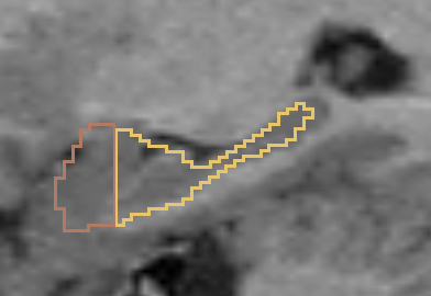
| 01:55, 8 April 2010 | RobustStatisticsSegmentation usage3.png (file) |  |
94 KB | 3 | |
| 00:20, 29 April 2008 | CA initialization.png (file) |  |
111 KB | the initialization of cellular automata algorithm | 1 |
| 21:52, 12 January 2012 | AFibEndoSegAtlasAndRSS axial.png (file) |  |
112 KB | 1 | |
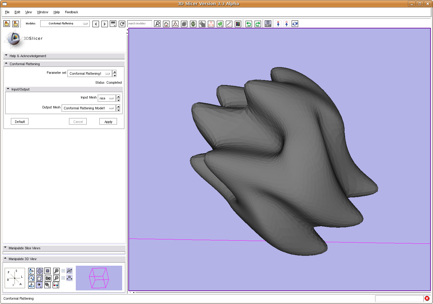
| 13:44, 27 June 2008 | SlicerConformalFlatten.png (file) |  |
126 KB | The surface we want to map to sphere. | 1 |
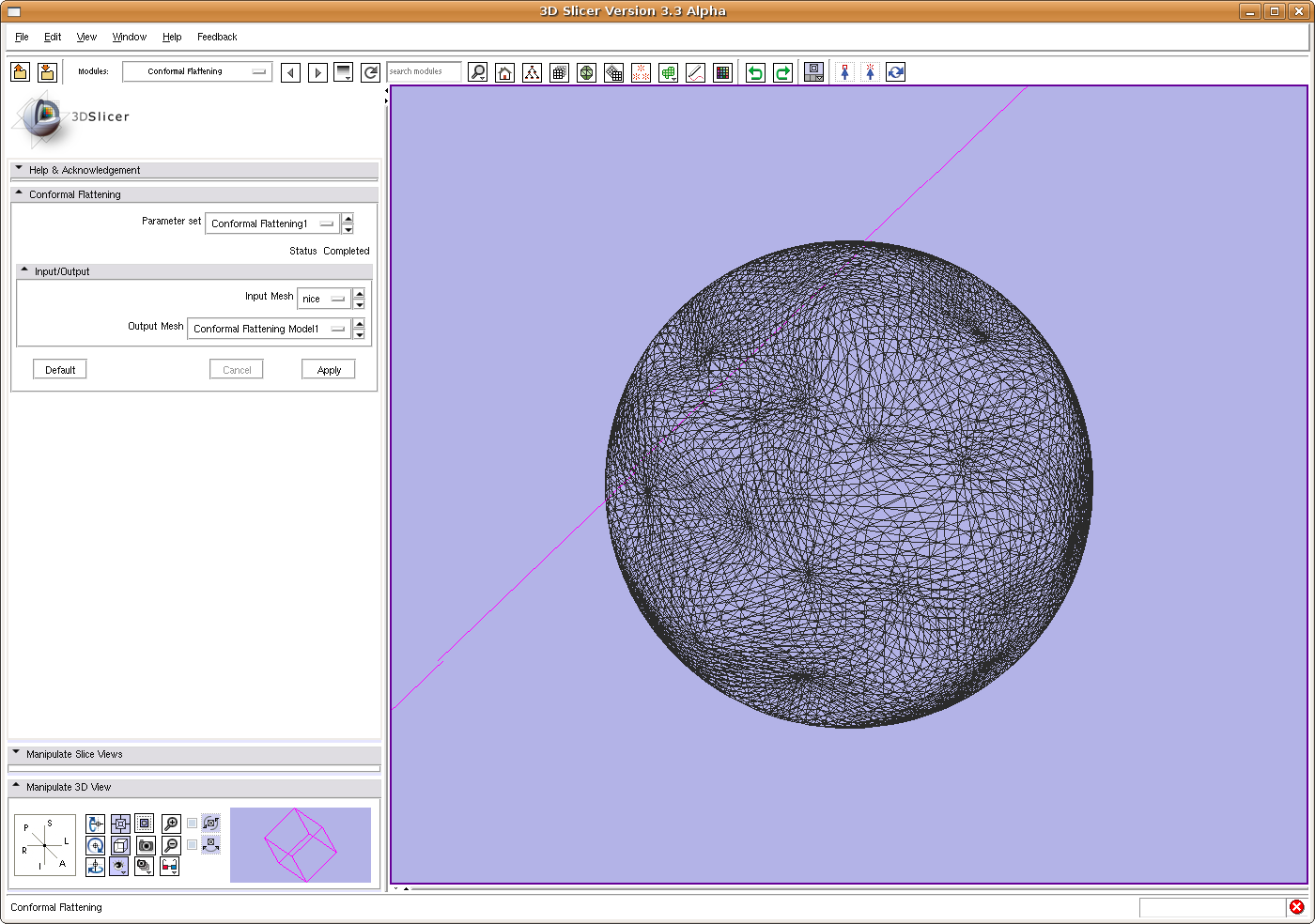
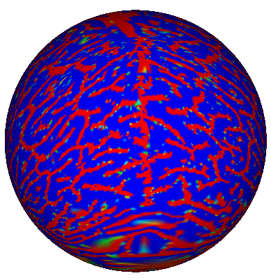
| 13:45, 27 June 2008 | SlicerConformalFlattenResult.png (file) |  |
135 KB | The mesh being mapped to sphere. | 1 |
| 03:51, 25 June 2010 | Hippo480-3d.png (file) |  |
144 KB | 1 | |
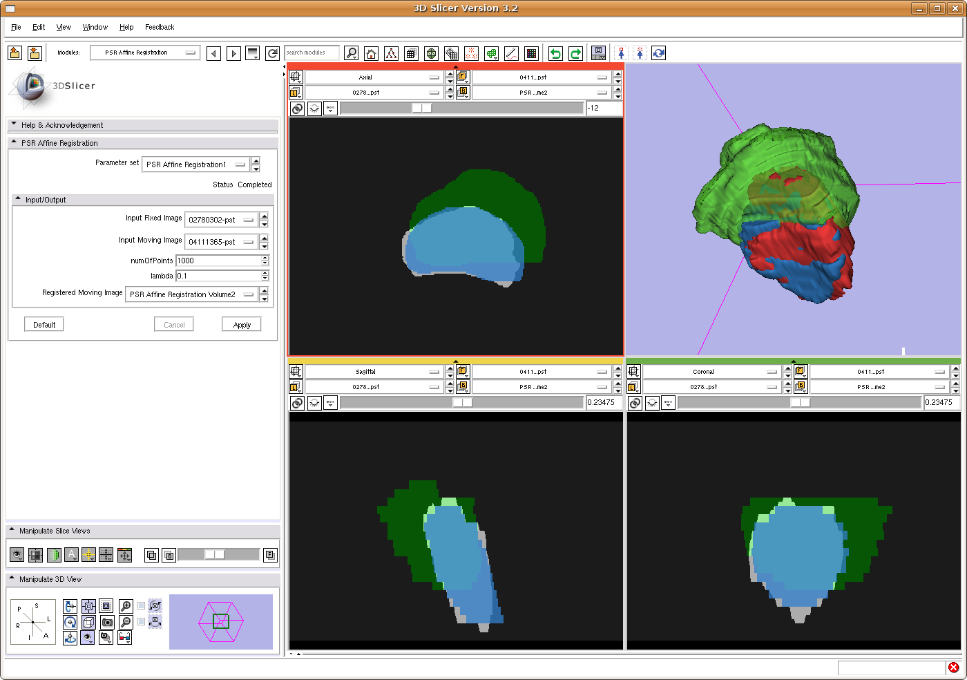
| 15:01, 24 June 2009 | ProstateRegistrationUsingPSRInSlicer.png (file) |  |
148 KB | Prostate affine registration in Slicer using the point set representation. Blue: Fixed image Green: Moving image Gray: Registered moving image In 3D view, registered moving image is in Red. | 1 |
| 19:19, 16 April 2007 | Brain-flat.PNG (file) |  |
152 KB | 1 | |
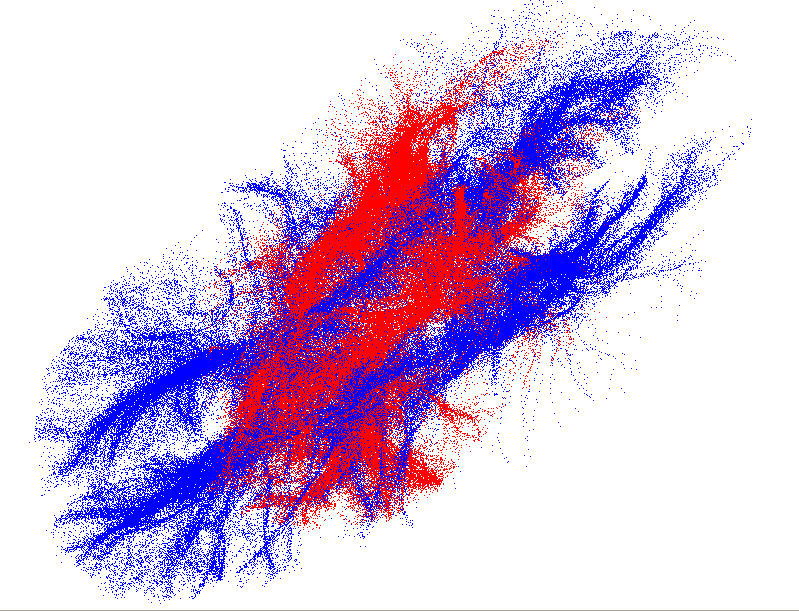
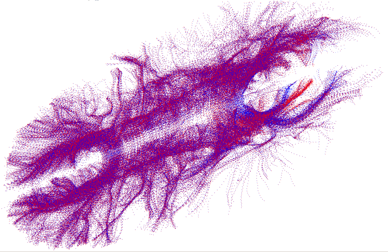
| 20:06, 12 April 2009 | BrainBefore.png (file) |  |
154 KB | The original brain point cloud in blue, and the affine transformed point cloud in red. The objective is to register the red to the blue. | 1 |
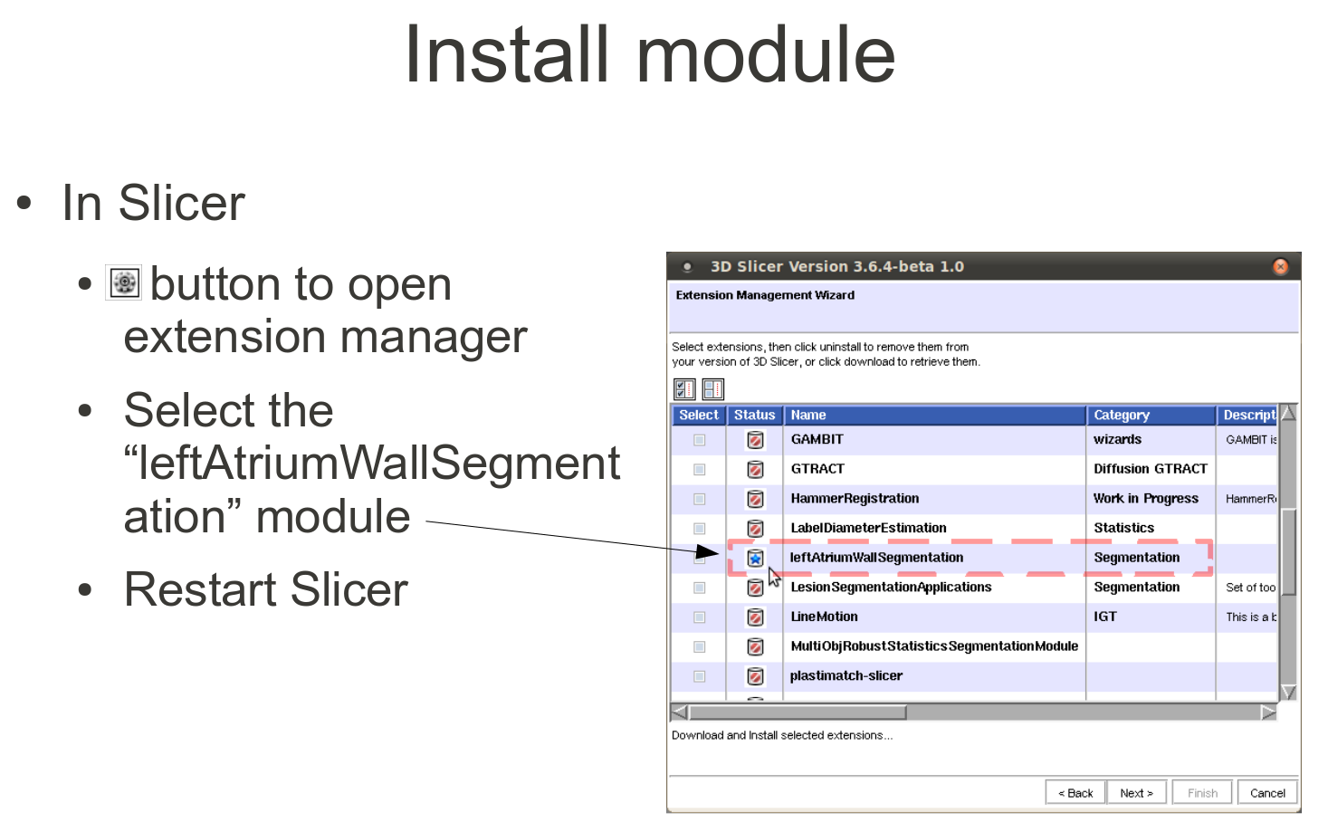
| 17:35, 21 March 2011 | WallSegmenterInstall.png (file) |  |
159 KB | 1 | |
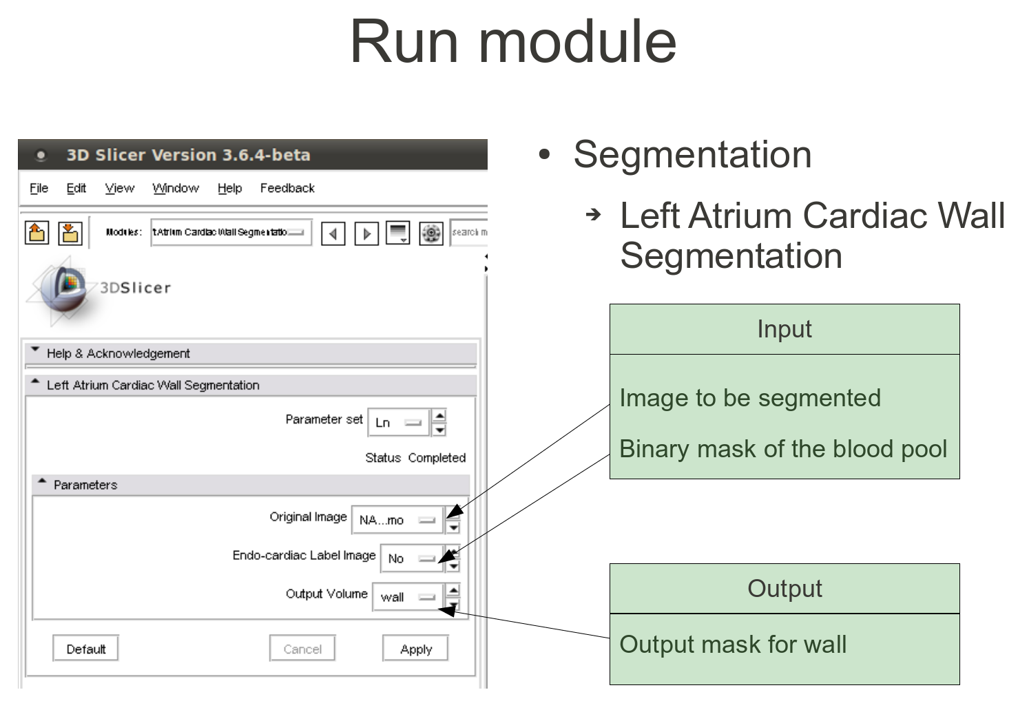
| 17:37, 21 March 2011 | WallSegmenterUse.png (file) |  |
161 KB | 1 | |
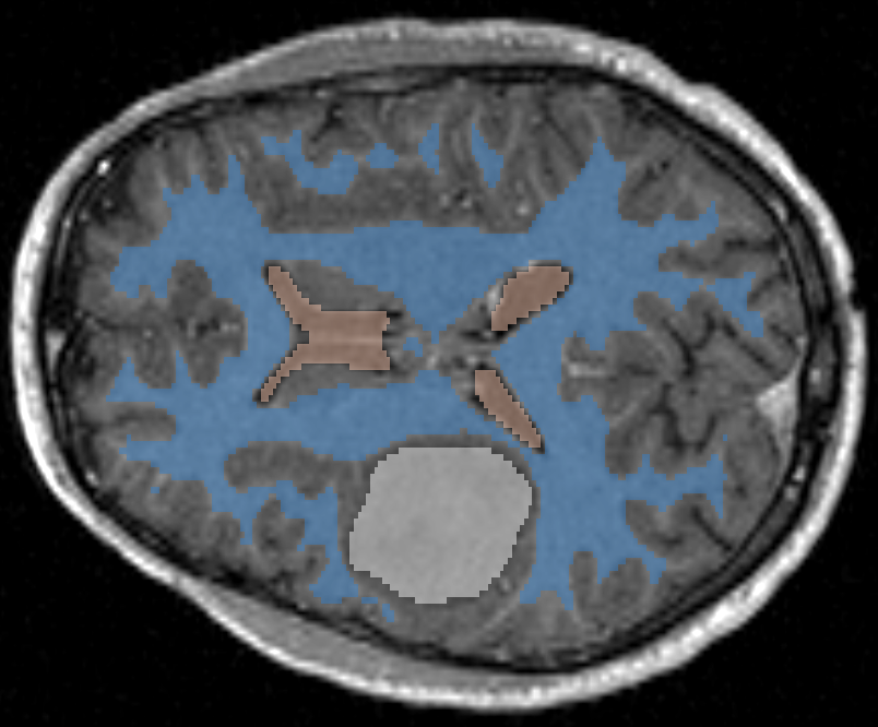
| 04:03, 8 January 2010 | MultiObjSeg.png (file) |  |
164 KB | 1 | |
| 20:07, 23 March 2009 | ProstateRegSupineToProneInParaview.png (file) |  |
172 KB | 2 | |
| 19:19, 16 April 2007 | Brain.PNG (file) |  |
178 KB | 1 | |
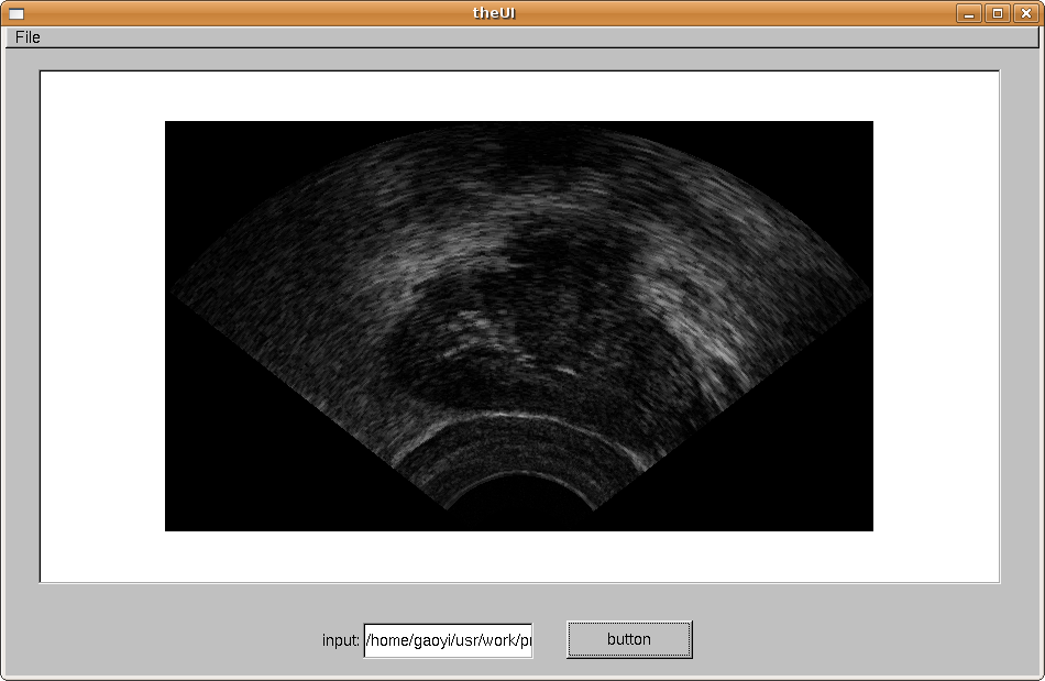
| 15:52, 11 January 2008 | UI prostate.png (file) |  |
180 KB | write a UI for the algorithm to interact with doctor. | 1 |
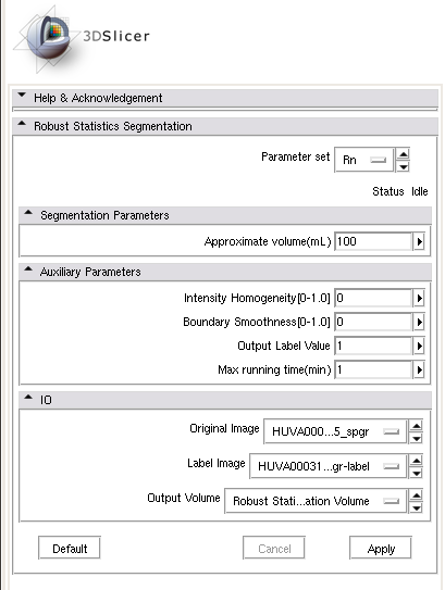
| 00:36, 8 April 2010 | RobustStatisticsSegmentation usage1.png (file) |  |
193 KB | 2 | |
| 22:58, 6 May 2010 | SPIE MI Gholami.pdf (file) | 193 KB | @conference{gao2010segmentation, title={{Segmentation of the Endocardial Wall of the Left Atrium using Local Region-Based Active Contours and Statistical Shape Learning}}, author={Gao, Y. and Gholami, B. and MacLeod, R.S. and Blauer, J. and Haddad, W. | 1 | |
| 22:43, 13 January 2011 | LaSeg1.png (file) |  |
219 KB | 1 | |
| 16:45, 1 March 2011 | Morss label 3 crop.png (file) |  |
224 KB | 1 | |
| 04:05, 8 January 2010 | TouchingNoOverlapping.png (file) |  |
232 KB | 1 | |
| 16:45, 1 March 2011 | Morss label 2 crop.png (file) |  |
237 KB | 1 | |
| 15:39, 1 March 2011 | RSS MultiObjSeg1.png (file) |  |
240 KB | 1 | |
| 04:04, 8 January 2010 | MultiObjSeg1.png (file) |  |
240 KB | 1 | |
| 20:06, 12 April 2009 | BrainAfter.png (file) |  |
248 KB | Registered Images Using Particle Filter Method. | 1 |
| 16:43, 1 March 2011 | Morss label 1 crop.png (file) |  |
256 KB | 1 | |
| 16:49, 1 March 2011 | Morss seg1.png (file) |  |
256 KB | 1 | |
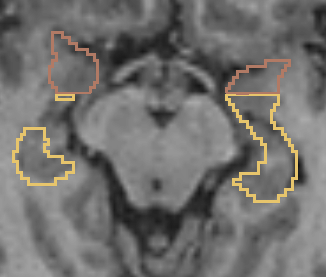
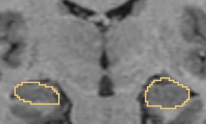
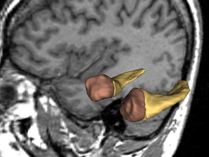
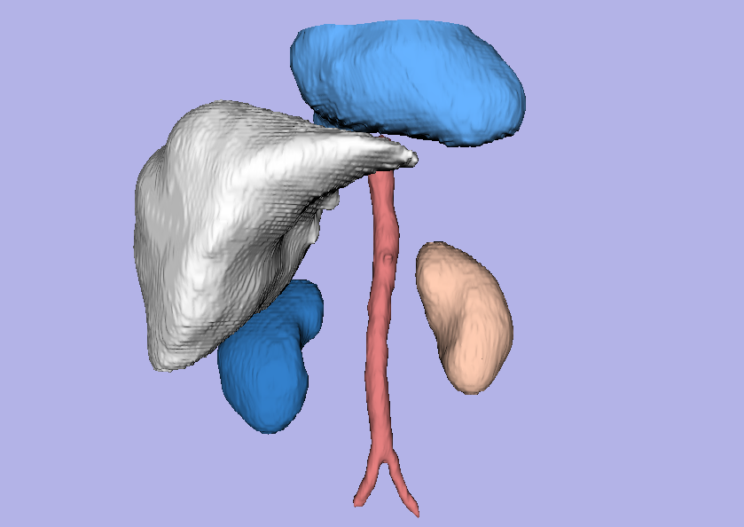
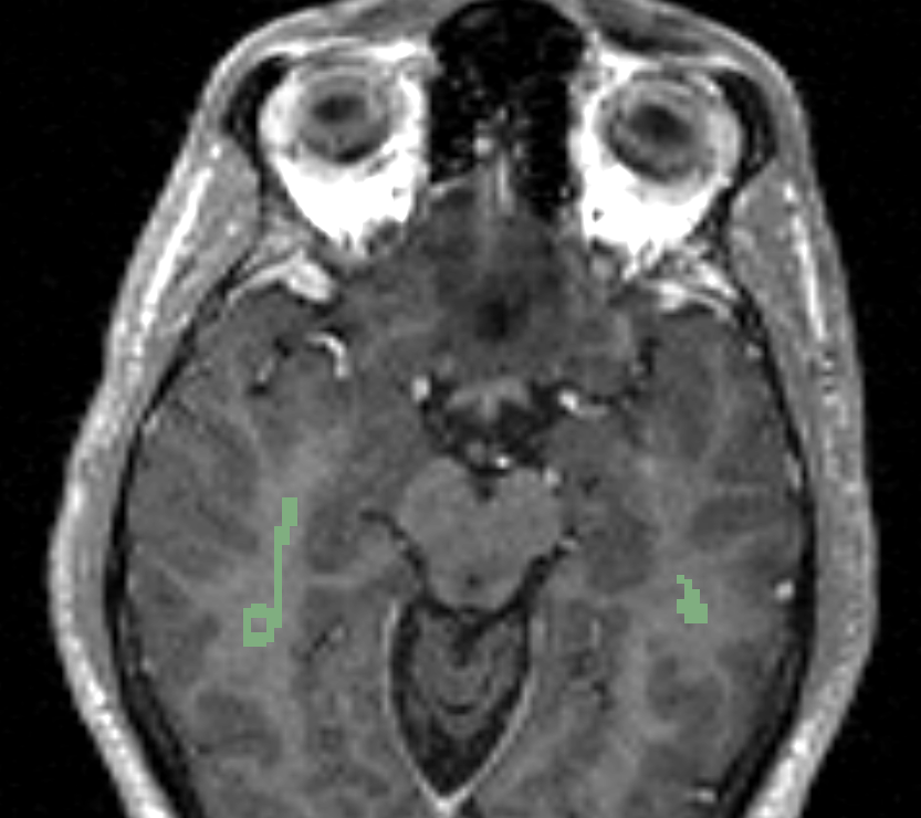
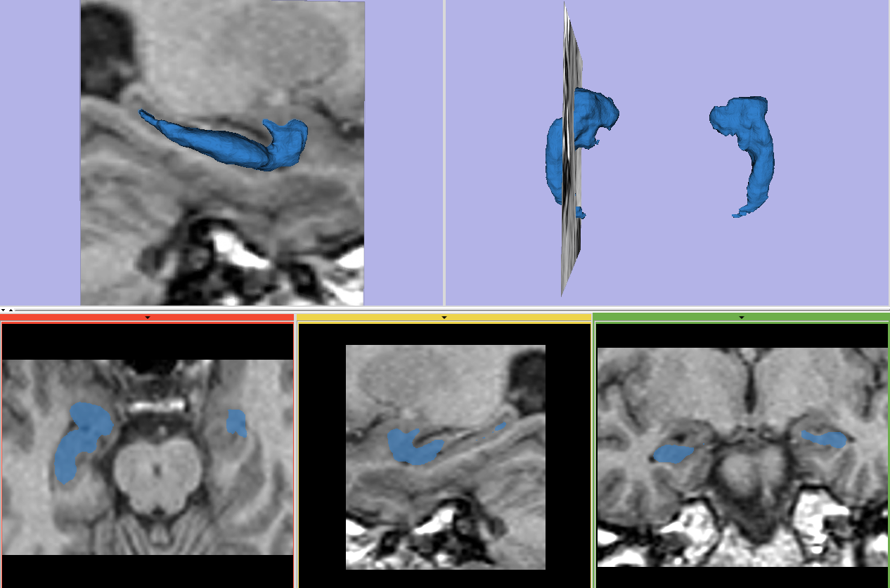
| 14:52, 11 May 2010 | SegHippoAmy.png (file) |  |
313 KB | Segmentation of hippocampus and amygdala | 1 |
| 16:49, 1 March 2011 | Morss seg2.png (file) |  |
326 KB | 1 | |
| 17:38, 21 March 2011 | LAwallSegmenterResult.png (file) |  |
333 KB | 1 | |
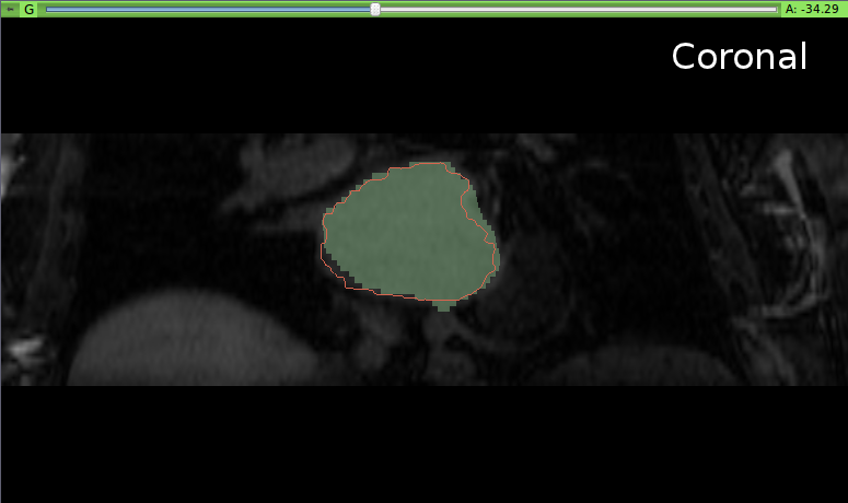
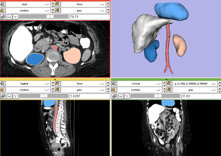
| 02:30, 24 June 2011 | RSSInEditorModule.png (file) |  |
338 KB | 1 |