Difference between revisions of "Stanford SIMBIOS: Whole Body Segmentation for Simulation"
(New page: ===Key Investigators=== * Stanford: Harish Doddi, Saikat Pal, and Scott Delp * WashU: Daniel Marcus * Harvard: ?? ===Tutorial===) |
|||
| (14 intermediate revisions by 2 users not shown) | |||
| Line 1: | Line 1: | ||
| + | {| | ||
| + | |[[Image:NAMIC-SLC.jpg|thumb|320px|Return to [[2009_Winter_Project_Week|Project Week Main Page]] ]] | ||
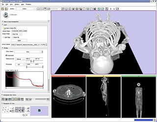
| + | |[[Image:ct.jpg|thumb|320px|Example whole-body CT dataset from WashU displayed in 3D Slicer.]] | ||
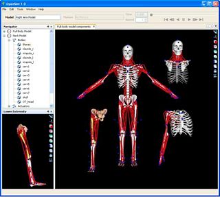
| + | |[[Image:sim.jpg|thumb|320px|Articulated musculoskeletal models.]] | ||
| + | |} | ||
===Key Investigators=== | ===Key Investigators=== | ||
| − | * Stanford: Harish Doddi, Saikat Pal | + | * Stanford: Harish Doddi, Saikat Pal |
* WashU: Daniel Marcus | * WashU: Daniel Marcus | ||
| − | * Harvard: | + | * Harvard: Ron Kikinis |
| + | * Steve Pieper, Isomics, Inc. | ||
| − | === | + | ===Project=== |
| + | __NOTOC__ | ||
| + | <div style="margin: 20px;"> | ||
| + | <div style="width: 40%; float: left; padding-right: 3%;"> | ||
| + | <h1>Objective</h1> | ||
| + | The aim of this project is to develop an automatic/semi-automatic methodology to convert whole body imaging datasets into three-dimensional models for neuromuscular biomechanics and finite element simulations. Initially, we will investigate the existing capabilities in EMSegmenter software to automatically segment the knee joint. | ||
| + | </div> | ||
| + | |||
| + | <div style="width: 27%; float: left; padding-right: 3%;"> | ||
| + | <h1>Approach, Plan</h1> | ||
| + | Investigate the existing functionality of EMSegementer to extract whole body models from CT and MR datasets. Initial efforts will be focused on developing atlases of specific joints (e.g. the knee) and evaluating EMSegmenter algorithms. The plan is to have imported MRI knee geometries in EMSegmenter and create an average atlas before the project week. During the project week, the EMSegmenter algorithm will be tested on a specific subject geometries. | ||
| + | </div> | ||
| + | |||
| + | <div style="width: 27%; float: left;"> | ||
| + | <h1>Progress</h1> | ||
| + | * Compiled a set of 5 knee MRI data-sets with manually segmented volumes. | ||
| + | * Evaluated registration algorithms to develop an averaged atlas of the knee joint. We evaluated the rigid and affine registrations techniques in Slicer, and attempted a diffeomorphic deamons algorithm to align multiple data-sets. | ||
| + | * Evaluated EMSegmenter to create atlas-independent image segmentation. | ||
| + | * We are in the process of converting manually-segmented models to a knee atlas. | ||
| + | |||
| + | </div> | ||
| + | |||
| + | <br style="clear: both;" /> | ||
| + | </div> | ||
Latest revision as of 17:11, 9 January 2009
Home < Stanford SIMBIOS: Whole Body Segmentation for Simulation Return to Project Week Main Page |
Key Investigators
- Stanford: Harish Doddi, Saikat Pal
- WashU: Daniel Marcus
- Harvard: Ron Kikinis
- Steve Pieper, Isomics, Inc.
Project
Objective
The aim of this project is to develop an automatic/semi-automatic methodology to convert whole body imaging datasets into three-dimensional models for neuromuscular biomechanics and finite element simulations. Initially, we will investigate the existing capabilities in EMSegmenter software to automatically segment the knee joint.
Approach, Plan
Investigate the existing functionality of EMSegementer to extract whole body models from CT and MR datasets. Initial efforts will be focused on developing atlases of specific joints (e.g. the knee) and evaluating EMSegmenter algorithms. The plan is to have imported MRI knee geometries in EMSegmenter and create an average atlas before the project week. During the project week, the EMSegmenter algorithm will be tested on a specific subject geometries.
Progress
- Compiled a set of 5 knee MRI data-sets with manually segmented volumes.
- Evaluated registration algorithms to develop an averaged atlas of the knee joint. We evaluated the rigid and affine registrations techniques in Slicer, and attempted a diffeomorphic deamons algorithm to align multiple data-sets.
- Evaluated EMSegmenter to create atlas-independent image segmentation.
- We are in the process of converting manually-segmented models to a knee atlas.

